Cardiomegaly: Difference between revisions
| Line 210: | Line 210: | ||
*:* Lateral Projection: The adult heart is 6 cm in the Antero Posterior (AP) direction | *:* Lateral Projection: The adult heart is 6 cm in the Antero Posterior (AP) direction | ||
[[image:cardiomegaly-2.jpg|left|400px|thumb|Cardiomegaly. <br> <small>Image courtesy of [[C. Michael Gibson]] MS. MD</small>]] | |||
<br clear="left"/> | |||
Image:Cardiomegaly-prosthetic-mitral-valve-001.jpg|left|400px|thumb|Cardiomegaly in a patients after mitral valve replacement. AP view. <br><small>Image courtesy of RadsWiki</small>]] | |||
<br clear="left"/> | |||
Image:Cardiomegaly-prosthetic-mitral-valve-001.jpg|Cardiomegaly in a patients after mitral valve replacement. AP view. <br><small>Image courtesy of RadsWiki | [[Image:Cardiomegaly-prosthetic-mitral-valve-002.jpg|left|400px|thumb|Cardiomegaly in a patients after mitral valve replacement. Lateral view. <br> <small>Image courtesy of RadsWiki</small>]] | ||
Image:Cardiomegaly-prosthetic-mitral-valve-002.jpg|Cardiomegaly in a patients after mitral valve replacement. Lateral view. <br> <small>Image courtesy of RadsWiki | <br clear="left"/> | ||
</ | |||
====X-ray findings for left atrial enlargement==== | ====X-ray findings for left atrial enlargement==== | ||
Revision as of 01:53, 28 February 2009
Template:DiseaseDisorder infobox
|
WikiDoc Resources for Cardiomegaly |
|
Articles |
|---|
|
Most recent articles on Cardiomegaly Most cited articles on Cardiomegaly |
|
Media |
|
Powerpoint slides on Cardiomegaly |
|
Evidence Based Medicine |
|
Clinical Trials |
|
Ongoing Trials on Cardiomegaly at Clinical Trials.gov Clinical Trials on Cardiomegaly at Google
|
|
Guidelines / Policies / Govt |
|
US National Guidelines Clearinghouse on Cardiomegaly
|
|
Books |
|
News |
|
Commentary |
|
Definitions |
|
Patient Resources / Community |
|
Patient resources on Cardiomegaly Discussion groups on Cardiomegaly Patient Handouts on Cardiomegaly Directions to Hospitals Treating Cardiomegaly Risk calculators and risk factors for Cardiomegaly
|
|
Healthcare Provider Resources |
|
Causes & Risk Factors for Cardiomegaly |
|
Continuing Medical Education (CME) |
|
International |
|
|
|
Business |
|
Experimental / Informatics |
| Cardiology Network |
 Discuss Cardiomegaly further in the WikiDoc Cardiology Network |
| Adult Congenital |
|---|
| Biomarkers |
| Cardiac Rehabilitation |
| Congestive Heart Failure |
| CT Angiography |
| Echocardiography |
| Electrophysiology |
| Cardiology General |
| Genetics |
| Health Economics |
| Hypertension |
| Interventional Cardiology |
| MRI |
| Nuclear Cardiology |
| Peripheral Arterial Disease |
| Prevention |
| Public Policy |
| Pulmonary Embolism |
| Stable Angina |
| Valvular Heart Disease |
| Vascular Medicine |
Template:WikiDoc Cardiology News Editor-In-Chief: C. Michael Gibson, M.S., M.D. [1]
Associate Editor in Chief: Cafer Zorkun, M.D., Ph.D. [2]
Please Take Over This Page and Apply to be Editor-In-Chief for this topic: There can be one or more than one Editor-In-Chief. You may also apply to be an Associate Editor-In-Chief of one of the subtopics below. Please mail us [3] to indicate your interest in serving either as an Editor-In-Chief of the entire topic or as an Associate Editor-In-Chief for a subtopic. Please be sure to attach your CV and or biographical sketch.
Overview
- Cardiomegaly is defined as an enlargement of the heart above its normal size.
- Cardiomegaly is generally first detected on chest X ray, and less often it is detected on routine physical examination.
- Cardiomegaly may be the first sign of an occult systemic or cardiovascular disease.
Pathophysiology
The left ventricle can be enlarged from two broad underlying conditions: dilation and hypertrophy.
- Left ventricular dilation:
- Can occurs as a result of volume overload. Conditions that cause volume overload can be further broken down as follows:
- Valvular Disease: More common underlying valvular heart disease conditions would include mitral regurgitation and aortic regurgitation
- Congenital Heart Disease: Patent ductus arteriosus and a ventricular septal defect.
- High output states resulting in volume overload: Anemia and thyrotoxicosis.
- High stroke volume states: Complete heart block and prolonged severe sinus bradycardia
- Can occur as a result of ischemia and remodeling
Differential Diagnosis of Cardiomegaly
In alphabetical order. [1] [2]
- Acromegaly
- Alcoholic cardiomyopathy
- Amyloidosis
- Anemia
- Atrial myxoma
- Carcinoid
- Chronic Obstructive Pulmonary Disease (COPD)
- Congenital heart disease
- Congestive Heart Failure CHF
- Cor pulmonale
- Drugs
- Endocardial fibroelastosis
- Epicardial fat pad
- Fabry's Disease
- Hemochromatosis
- Hurler's Syndrome
- Hyperparathyroidism
- Hypertension
- Hypertrophic cardiomyopathy
- Hypocalcemia
- Hypomagnesemia
- Hypoparathyroidism
- Hypophosphatemia
- Idiopathic cardiomyopathy
- Ischemic heart disease
- Kyphoscoliosis
- Marfan's Syndrome
- Mediastinal mass
- Myocarditis
- Normal, "athletic" heart
- Pompe's Disease
- Postradiation
- Pregnancy
- Primary pulmonary hypertension
- Pulmonary embolism
- Renal failure: Acute Renal Failure and Chronic Renal Failure
- Scleroderma
- Sickle cell disease
- Subacute bacterial endocarditis
- Systemic Lupus Erythematosus
- Valvular heart disease
Differential Diagnosis of Specific Chamber Enlargements
Left Atrial Enlargement
- Atrial aneurysm
- Infective endocarditis
- Left heart failure
- Mitral regurgitation
- Mitral stenosis
- Mitral valve prolapse
- Myxedma
- Patent Ductus Arteriosus
- Ventricular septal defect
Left Ventricular Hypertrophy
- Acromegaly
- Alcoholic cardiomyopathy
- Aortic coarctation
- Aortic regurgitation
- Aortic stenosis
- Chronic Renal Failure
- Familial dilated and hypertrophic cardiomyopathy
- Hypertrophic cardiomyopathy (HCM)
- Idiopathic dilated cardiomyopathy
- Mitral regurgitation
- Patent Ductus Arteriosus
- Restrictive cardiomyopathy
- Thalassemia
Right Atrial Enlargement
- Atrial aneurysm
- Atrial septal defect
- Ebstein's anomaly
- Pulmonic stenosis
- Right heart failure
- Tricuspid prolapse
- Tricuspid regurgitation
- Tricuspid stenosis
- Tumor
Right Ventricular Hypertrophy
- Atrial septal defect
- Chronic Obstructive Pulmonary Disease (COPD)
- Cor pulmonale
- Eisenmenger's Syndrome
- Left ventricular hypertrophy or Left ventricular dilation
- Mitral stenosis
- Pickwickian Syndrome
- Pulmonic regurgitation
- Pulmonic stenosis
- Tetralogy of Fallot
- Transposition of the Great Vessels
- Tricuspid regurgitation
- Ventricular septal defect
Diagnosis
History and Symptoms
- In the history, the physician should inquire as to:
- History of chest pain to suggest ischemic heart disease
- History of rheumatic fever or known valvular heart disease
- History of congenital heart disease
- History of drug and alcohol use (recreational)
- Family history (heart disease or sudden death)
- Associated symptoms:
Physical Examination
Heart
There is downward and leftward displacement of the heart on percussion of heart border. The heart should be percussed with the patient supine and the head of the bed elevated 30 degrees. Normally the left heart border should not
- Extend left of the mid clavicular line
- Extend more than 10 cm to the left of the mid sternal line
- Occupy a diameter > 3 cm
- Extend below the 5th intercostal space
The body habitus of the individual must be taken into account in performing the examination. In athletic individuals who are thin, the impulse maybe strongly transmitted. In muscular or obese patients, the opposite is true. In patients with pectus excavatum, the apex can be displaced laterally.
Laboratory Findings
Based upon the history and physical, a standard evaluation might include:
Common high yield tests:
- Complete blood count (CBC): to rule out anemia
- Thyroid stimulating hormone (TSH)
- Blood urea nitrogen (BUN) / creatinine
Less frequent low yield tests:
- Erythrocyte sedimentation rate (ESR): to evaluate autoimmune causes
- Calcium: to rule out hypocalcemia
- Magnesium: to rule out hypomagnesemia
- Rheumatoid factor
- Antinuclear antibody (ANA)
- Glucose
- Pheochromocytoma screening
- Phosphorus: to rule out hypophosphatemia
- Blood cultures
Electrolyte and Biomarker Studies
- Electrolytes should be periodically checked in the patient treated with a diuretic to avoid hypokalemia.
- Brain Natriuretic Peptide (BNP) levels may provide insight into the ongoing magnitude of LV dysfunction.
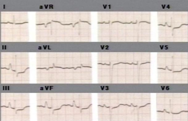
Electrocardiogram
- The presence of q waves suggest a prior MI to account for the enlargement.
- ST segment elevation would suggest either ongoing or resolving ST Elevation Myocardial Infarction or Myocarditis or Pericarditis.
- The EKG may suggest signs of left atrial enlargement, right atrial enlargement or right axis deviation which may point toward a specific diagnosis.
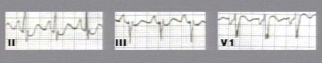
Images shown below are courtesy of Professor Peter Anderson DVM PhD and published with permission. © PEIR, University of Alabama at Birmingham, Department of Pathology
-
Right atrial enlargement
-
Biatrial enlargement

Chest X Ray
- Cardiomgaly is traditionally defined as an increase in the cardiothoracic ratio to be > 0.5 on a PA film. To calculate the thoracic ratio, the width of the cardiac silhouette is divided by the width of the entire thoracic cage.
- If the heart is viewed on an AP film, the heart can appear to be artificially enlarged because the X ray beam moves from anterior to posterior direction and therefore the heart which lies anterior is magnified.
- Postero Anterior (PA) Projection: adult heart is 12 cm from base to apex and 8-9 cm in transverse direction
- Lateral Projection: The adult heart is 6 cm in the Antero Posterior (AP) direction

Image courtesy of C. Michael Gibson MS. MD
Image:Cardiomegaly-prosthetic-mitral-valve-001.jpg|left|400px|thumb|Cardiomegaly in a patients after mitral valve replacement. AP view.
Image courtesy of RadsWiki]]

Image courtesy of RadsWiki
X-ray findings for left atrial enlargement
- Double density sign: Occur when the right side of the left atrium pushes into the adjacent lung.
- Convex left atria appendage: usually reflect prior rheumatic heart disease
- Splaying of the carina
- Posterior displacement of the left main stem bronchus on lateral radiograph
- Superior displacement of the left main stem bronchus on frontal view
- Posterior displacement of a barium filled esophagus


X-ray findings for right ventricular enlargement
- Frontal view
- Rounded left heart border
- Uplifted apex
- Lateral view
- Filling of the retrosternal space
- Rotation of the heart posteriorly
X-ray findings for right atrial enlargement
- On a frontal view, the right atrium is visible because of its interface with the right middle lobe.
- Subtle and moderate right atrial enlargement is not accurately determined on plain films because there is normal variability in the shape of the right atrium.
Echocardiography or Ultrasound
- Echocardiogram recommended for those patients presenting suspected valvular disease, chamber size, ventricular function, and wall motion abnormalities
Other Diagnostic Studies
- Exercise or pharmacologic stress testing may be indicated in those patients suspected of having coronary artery disease
- Patients suspected of having valvular disease or coronary artery disease may need a cardiac catheterization for full evaluation following echocardiography
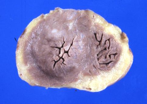
Pathologic Findings
Image shown below is courtesy of Professor Peter Anderson DVM PhD and published with permission. © PEIR, University of Alabama at Birmingham, Department of Pathology
-

Right ventricular enlargement due to a patent ductus arteriosis in a patient with hyaline membrane disease

Treatment
Pharmacotherapy
Acute Pharmacotherapies
- Administration of digoxin, diuretics, antiarrhythmics and/or preload and afterload reducers per clinical indication
Chronic Pharmacotherapies
A combination of diuretics and angiotensin converting enzyme (ACE) inhibition is currently the standard of care. Digoxin may reduce the frequency of rehospitalization, but does not improve mortality.
Surgery and Device Based Therapy
- In patients who are awaiting a transplant for end-stage symptomatic heart failure, implantable ventricular assist devices may serve as a temporary aid for compensation.
Transplantation
- In patients who are in end-stage symptomatic heart failure, a heart transplant my be necessary.
Future or Investigational Therapies
Mechanical strategies currently under investigation include:
- Development of devices to reduce the size of the heart
- Development of a device to exclude that portion of the apex where clots may form.
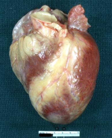
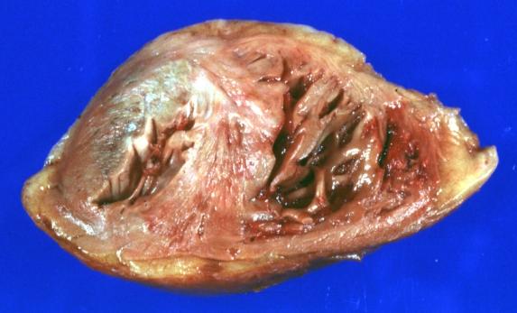
Pathological Findings
Images shown below are courtesy of Professor Peter Anderson DVM PhD and published with permission. © PEIR, University of Alabama at Birmingham, Department of Pathology
-
Biventricular Hypertrophy
-

Biventricular Hypertrophy


-
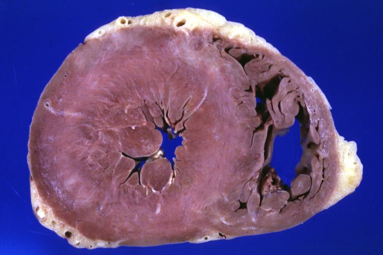
Gross excellent example of concentric left ventricular hypertrophy
-
Left Ventricular Hypertrophy: Gross natural color anterior view intact heart showing disproportionate size of left ventricle by its inferior extent much below the right ventricle apex (quite good example)


-
Myocardial Infarct: Gross natural color apical section showing large left ventricle infarct and right ventricular hypertrophy
-

Right ventricular hypertrophy


References
Additional Reading
- Moss and Adams' Heart Disease in Infants, Children, and Adolescents Hugh D. Allen, Arthur J. Moss, David J. Driscoll, Forrest H. Adams, Timothy F. Feltes, Robert E. Shaddy, 2007 ISBN 0781786843
- Hurst's the Heart, Fuster V, 12th ed. 2008, ISBN 978-0-07-149928-6
- Willerson JT, Cardiovascular Medicine, 3rd ed., 2007, ISBN 978-1-84628-188-4
Acknowledgements
The content on this page was first contributed by C. Michael Gibson, M.S., M.D.