Appendix cancer pathophysiology
|
Appendix cancer Microchapters |
|
Diagnosis |
|---|
|
Treatment |
|
Appendix cancer pathophysiology On the Web |
|
American Roentgen Ray Society Images of Appendix cancer pathophysiology |
|
Risk calculators and risk factors for Appendix cancer pathophysiology |
Editor-In-Chief: C. Michael Gibson, M.S., M.D. [1]; Associate Editor(s)-in-Chief: Soroush Seifirad, M.D.[2]
Overview
The pathophysiology of appendix cancer depends on the histological subtype. There are two major subtypes of appendix cancer, adenocarcinomas and carcionid tumors. While carcinoid tumors arises from enterochromaffin cells (Kulchitsky cells), which are secretory cells that are normally involved in neuroendocrine hormonal secretions, adenocarcinomas are the result of mutations in mucous producing epithelial cells. Their physiology, pathophysiology, genetic pathways, prognosis as well as epidemiology are different and hence, discussed separately. The progression to adenocarcinoma usually involves the KRAS, APC, TP53, and RAF pathways, While β-catenin, NF1, and MEN1 genes are major contributors of carcinoid tumors progression.
Pathophysiology
Physiology
- The normal physiology of entrochromaffin cells is secretion of serotonin (5-HT), histamine, kallikrein, prostaglandins, and tachykinins.
- Glandular epithelial cells are responsible for mucous production.
Pathogenesis
- The pathophysiology of appendix cancer depends on the histological subtype.
- Adenocarcinoma arises from epithelial glandular cells, which are normally involved in mucous production.
- Carcinoid tumors arise from entrochromaffin cell, which are neuroendocrine cells that are normally involved in secretion of serotonin (5-HT), histamine, kallikrein, prostaglandins, and tachykinins.
- The pathogenesis of appendix cancer is characterized by an initial epithelial dysplasia, followed by the formation of cystic structures and angiolymphatic invasion. Subsequently, in the advanced stages of appendix cancer, tumor cells detach from the primary tumor mass and gain access to the peritoneal cavity.[1]
Genetics
Genes involved in the pathogenesis of carcinoid tumors of appendix include:
The development of appendiceal adeocarcinoma is the result of multiple genetic mutations such as:
Associated Conditions
- Conditions associated with appendiceal cancers include:
- Chronic inflammatory disease such as Ulcerative colitis
- Familial cancer syndromes
Gross Pathology
- On gross pathology, findings of appendix cancer, include:[1]
- Well-demarcated mass
- Average size between 1 and 5 cm
- Gray or yellowish color
- Deformed appendix
- Adenocarcinoma
- Gray/yellow color
- Cystic structures with angiolymphatic invasion
- Appendix might be buried within the mass
- Carcinoid tumors
- Prevalent at the tip of appendix
- Generally less than 1 cm
- Gray or yellow
- Well-demarcated firm
- Intramural nodules that may narrow or obliterate appendiceal lumen
- Proximal tumors may cause obstruction and appendicitis
- Goblet cell carcinoids
- No gross tumor might be present
- Thickened appendiceal wall
- The image below demonstrates gross pathology of appendix cancer.
-
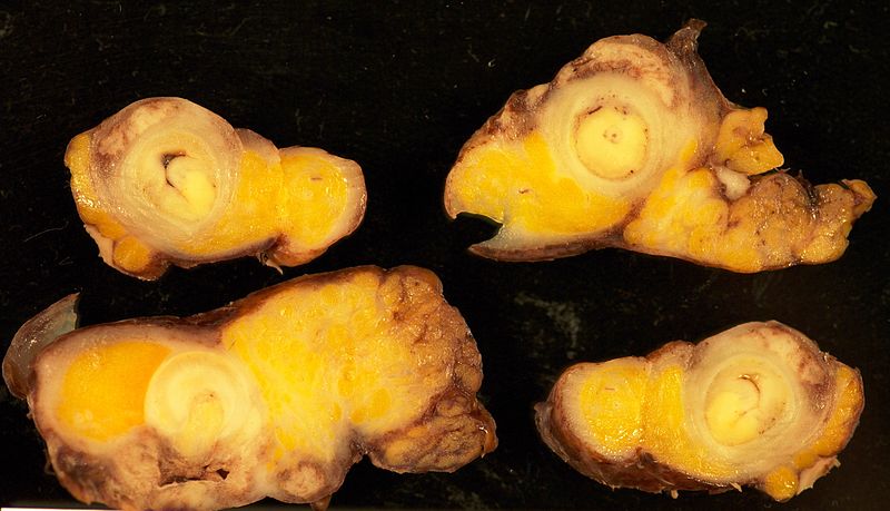
Gross pathology appendix cancer

Microscopic Pathology
- On microscopic histopathological analysis findings will depend on the subtype of appendicular cancer.
- Common histopathological findings, may include:[1]
- Cystic structures
- Angiolymphatic invasion
- Adenocarcinoma
- Intestinal, mucinous or signet ring cell types
- oexisting acute appendicitis is common IHC might be positive for the following stains:
- MUC 2
- MUC5AC
- CK 8/18
- CK 13
- CK 19
- CK 20
- Carcinoid tumor
- Insular growth pattern of solid islands of uniform polygonal cells with minimal pleomorphism
- Retraction of peripheral tumor cells from stroma
- Angiolymphatic invasion is common
- Granular eosinophilic cytoplasm with either diffusely scattered or peripherally clumped granules
- Two types of well differentiated tumors: EC cell (serotonin producing) and rarely L-cell (enteroglucagon or peptide YY producing) IHC might be positive for S100
- Goblet cell
- GCC Generally spares mucosa and infiltrates muscularis propria and periappendiceal fat
- Tumor cell clusters
- Crypt-like structures
- Tubules of mucus-secreting cells distended with mucin resembling goblet cells
- Eosinophilic cytoplasm resembling carcinoid tumors
- Pools of extracellular mucin
- Scattered Paneth cells in tumors with crypt like structures
- Extensive perineural invasion
- Carcinomatous growth pattern:
- Cribriform growth pattern, solid sheets of infiltrating signet ring cells
- Nuclear pleomorphism
- Increased mitotic activity
- The images below demonstrate different histopathological findings of appendix cancer.
-
![Appendiceal carcinoid[2]](/images/2/20/800px-Appendix_Carcinoid_Torsion_1X_PA.JPG)
Appendiceal carcinoid[2]
-
![Appendiceal carcinoid[2]](/images/e/e1/800px-Appendix_Carcinoid_HP_14BR---.jpg)
Appendiceal carcinoid[2]
-
![Appendiceal carcinoid with necrosis[2]](/images/6/68/800px-Appendix_Carcinoid_Necrosis_PA.JPG)
Appendiceal carcinoid with necrosis[2]
-
![Carcinoid synaptophysin[2]](/images/a/a3/800px-Appendix_Carcinoid_Synaptophysin_14BR---.jpg)
Carcinoid synaptophysin[2]
-
![Appendiceal tumor[2]](/images/2/28/800px-Appendix_Carcinoid_HP_CTR.jpg)
Appendiceal tumor[2]
![Appendiceal carcinoid[2]](/index.php/File:800px-Appendix_Carcinoid_Torsion_1X_PA.JPG)
![Appendiceal carcinoid[2]](/index.php/File:800px-Appendix_Carcinoid_HP_14BR---.jpg)
![Appendiceal carcinoid with necrosis[2]](/index.php/File:800px-Appendix_Carcinoid_Necrosis_PA.JPG)
![Carcinoid synaptophysin[2]](/index.php/File:800px-Appendix_Carcinoid_Synaptophysin_14BR---.jpg)
![Appendiceal tumor[2]](/index.php/File:800px-Appendix_Carcinoid_HP_CTR.jpg)
References
- ↑ 1.0 1.1 1.2 Ruoff C, Hanna L, Zhi W, Shahzad G, Gotlieb V, Saif MW (2011). "Cancers of the appendix: review of the literatures". ISRN Oncol. 2011: 728579. doi:10.5402/2011/728579. PMC 3200132. PMID 22084738.
- ↑ 2.0 2.1 2.2 2.3 2.4 http://librepathology.org/wiki/index.php/Neuroendocrine_tumour_of_the_appendix