Appendix cancer pathophysiology
|
Appendix cancer Microchapters |
|
Diagnosis |
|---|
|
Treatment |
|
Appendix cancer pathophysiology On the Web |
|
American Roentgen Ray Society Images of Appendix cancer pathophysiology |
|
Risk calculators and risk factors for Appendix cancer pathophysiology |
Editor-In-Chief: C. Michael Gibson, M.S., M.D. [1]; Associate Editor(s)-in-Chief: Soroush Seifirad, M.D.[2]
Overview
The pathophysiology of appendix cancer depends on the histological subtype. There are two major subtypes of appendix cancer, adenocarcinomae and carcionid tumors. While carcinoid tumors arises from enterochromaffin cells (Kulchitsky cells), which are secretory cells that are normally involved in neuroendocrine hormonal secretions, adenocarcinomae are the result of mutations in mucous producing epithelial cells. Their physiology, pathophysiology, genetic pathways, prognosis as well as epidemiology are different and hence discussed separately. The progression to adenocarcinoma usually involves the KRAS, APC, TP53, and RAF pathways, While β-catenin, NF1, and MEN1 genes are major contributors of carcinoid tumor s progression.
Pathophysiology
Physiology
The normal physiology of entrochromaffin cells is secretion of serotonin (5-HT), histamine, kallikrein, prostaglandins, and tachykinins.
Glandular epithelial cells are responsible for mucous production.
Pathogenesis
- The exact pathogenesis of [disease name] is not completely understood.
OR
- It is understood that [disease name] is the result of / is mediated by / is produced by / is caused by either [hypothesis 1], [hypothesis 2], or [hypothesis 3].
- [Pathogen name] is usually transmitted via the [transmission route] route to the human host.
- Following transmission/ingestion, the [pathogen] uses the [entry site] to invade the [cell name] cell.
- [Disease or malignancy name] arises from [cell name]s, which are [cell type] cells that are normally involved in [function of cells].
- The progression to [disease name] usually involves the [molecular pathway].
- The pathophysiology of [disease/malignancy] depends on the histological subtype.
Genetics
[Disease name] is transmitted in [mode of genetic transmission] pattern.
OR
Genes involved in the pathogenesis of [disease name] include:
- [Gene1]
- [Gene2]
- [Gene3]
OR
The development of [disease name] is the result of multiple genetic mutations such as:
- [Mutation 1]
- [Mutation 2]
- [Mutation 3]
Associated Conditions
Conditions associated with [disease name] include:
- [Condition 1]
- [Condition 2]
- [Condition 3]
Gross Pathology
On gross pathology, [feature1], [feature2], and [feature3] are characteristic findings of [disease name].
Microscopic Pathology
On microscopic histopathological analysis, [feature1], [feature2], and [feature3] are characteristic findings of [disease name].
References
Pathophysiology
- The pathogenesis of appendix cancer is characterized by an initial epithelial dysplasia, followed by the formation of cystic structures and angiolymphatic invasion. Subsequently, in the advanced stages of appendix cancer, tumor cells detach from the primary tumor mass and gain access to the peritoneal cavity.[1]
- The KRAS gene mutation has been associated with the development of appendix cancer.
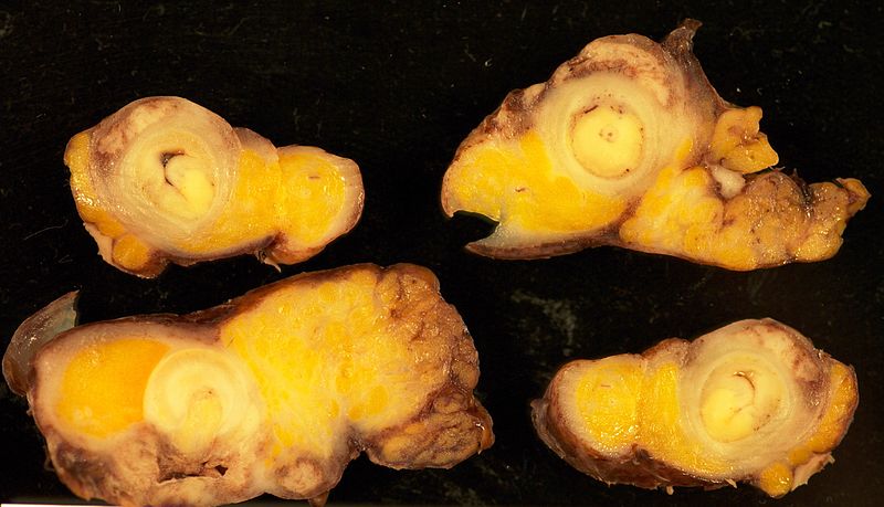
- On gross pathology, findings of appendix cancer, include:[1]
- Well-demarcated mass
- Average size between 1 and 5 cm
- Gray or yellowish color
- Deformed appendix
- The image below demonstrates gross pathology of appendix cancer.
-
Gross pathology appendix cancer
- On microscopic histopathological analysis findings will depend on the subtype of appendicular cancer.
- Common histopathological findings, may include:[1]
- Cystic structures
- Angiolymphatic invasion
- The images below demonstrate different histopathological findings of appendix cancer.
-
Appendiceal carcinoid[2]
-
Appendiceal carcinoid[2]
-
Appendiceal carcinoid with necrosis[2]
-
Carcinoid synaptophysin[2]
-
Appendiceal tumor[2]
References
- ↑ 1.0 1.1 1.2 Ruoff C, Hanna L, Zhi W, Shahzad G, Gotlieb V, Saif MW (2011). "Cancers of the appendix: review of the literatures". ISRN Oncol. 2011: 728579. doi:10.5402/2011/728579. PMC 3200132. PMID 22084738.
- ↑ 2.0 2.1 2.2 2.3 2.4 http://librepathology.org/wiki/index.php/Neuroendocrine_tumour_of_the_appendix

![Appendiceal carcinoid[2]](/images/2/20/800px-Appendix_Carcinoid_Torsion_1X_PA.JPG)
![Appendiceal carcinoid[2]](/images/e/e1/800px-Appendix_Carcinoid_HP_14BR---.jpg)
![Appendiceal carcinoid with necrosis[2]](/images/6/68/800px-Appendix_Carcinoid_Necrosis_PA.JPG)
![Carcinoid synaptophysin[2]](/images/a/a3/800px-Appendix_Carcinoid_Synaptophysin_14BR---.jpg)
![Appendiceal tumor[2]](/images/2/28/800px-Appendix_Carcinoid_HP_CTR.jpg)