Appendix cancer CT scan: Difference between revisions
Jump to navigation
Jump to search
No edit summary |
No edit summary |
||
| Line 19: | Line 19: | ||
** Sometimes it is challenging to distinguish between tumor and mucin. | ** Sometimes it is challenging to distinguish between tumor and mucin. | ||
** There are reports in favor of Diffusion weighted MRI superiority compared to CT in evaluating extent of peritoneal involvement.<ref name="pmid22302265">Low RN, Barone RM (2012) [https://www.ncbi.nlm.nih.gov/entrez/eutils/elink.fcgi?dbfrom=pubmed&retmode=ref&cmd=prlinks&id=22302265 Combined diffusion-weighted and gadolinium-enhanced MRI can accurately predict the peritoneal cancer index preoperatively in patients being considered for cytoreductive surgical procedures.] ''Ann Surg Oncol'' 19 (5):1394-1401. [http://dx.doi.org/10.1245/s10434-012-2236-3 DOI:10.1245/s10434-012-2236-3] PMID: [https://pubmed.gov/22302265 22302265]</ref> | ** There are reports in favor of Diffusion weighted MRI superiority compared to CT in evaluating extent of peritoneal involvement.<ref name="pmid22302265">Low RN, Barone RM (2012) [https://www.ncbi.nlm.nih.gov/entrez/eutils/elink.fcgi?dbfrom=pubmed&retmode=ref&cmd=prlinks&id=22302265 Combined diffusion-weighted and gadolinium-enhanced MRI can accurately predict the peritoneal cancer index preoperatively in patients being considered for cytoreductive surgical procedures.] ''Ann Surg Oncol'' 19 (5):1394-1401. [http://dx.doi.org/10.1245/s10434-012-2236-3 DOI:10.1245/s10434-012-2236-3] PMID: [https://pubmed.gov/22302265 22302265]</ref> | ||
** | |||
{| class="wikitable" | |||
|+PCI Scoring System | |||
! colspan="2" |Lesion Size Score | |||
|- | |||
|LS0 | |||
|No tumor seen | |||
|- | |||
|LS1 | |||
|Tumor up to 0.5 cm | |||
|- | |||
|LS2 | |||
|Tumor up to 5 cm cm | |||
|- | |||
|LS3 | |||
|Tumor > 5 cm or confluence | |||
|- | |||
| colspan="2" |''Maximum Score = 3'' | |||
|- | |||
! colspan="2" |Regions (0-3) | |||
|- | |||
|0 | |||
|Central | |||
|- | |||
|1 | |||
|Right Upper | |||
|- | |||
|2 | |||
|Epigasterium | |||
|- | |||
|3 | |||
|Left Upper | |||
|- | |||
|4 | |||
|Left Flank | |||
|- | |||
|5 | |||
|Left Lower | |||
|- | |||
|6 | |||
|Pelvis | |||
|- | |||
|7 | |||
|Right Upper | |||
|- | |||
|8 | |||
|Right Flank | |||
|- | |||
|9 | |||
|Upper Jejunum | |||
|- | |||
|10 | |||
|Lower Jejunum | |||
|- | |||
|11 | |||
|Upper Illeum | |||
|- | |||
|12 | |||
|lower Illeum | |||
|- | |||
| colspan="2" |''Maximum Score = 36'' | |||
|- | |||
| colspan="2" |'''Total Maximum Score = 39''' | |||
|} | |||
**[[File:Peritoneal Carcinomatosis Index (PCI) Regions.jpg|thumb|'''Peritoneal Carcinomatosis Index (PCI) Regions''']] | |||
* CT scan also helps in discovering distant metastatic lessons in the other organs like bone, lungs, and brain. | * CT scan also helps in discovering distant metastatic lessons in the other organs like bone, lungs, and brain. | ||
Revision as of 18:58, 22 January 2019
|
Appendix cancer Microchapters |
|
Diagnosis |
|---|
|
Treatment |
|
Appendix cancer CT scan On the Web |
|
American Roentgen Ray Society Images of Appendix cancer CT scan |
|
Risk calculators and risk factors for Appendix cancer CT scan |
Editor-In-Chief: C. Michael Gibson, M.S., M.D. [1]; Associate Editor(s)-in-Chief:
Overview
Abdominal CT scan is pretty helpful in the diagnosis of appendix cancer. Findings on CT scan suggestive of appendix cancer include [finding 1], [finding 2], and [finding 3]. CT scan is one of the best imaging modalities to assess disease burden, metastatic lesions as well as disease stage.
CT scan
- Abdominal CT scan may be helpful in the diagnosis of appendiceal cancer. Findings on CT scan suggestive of appendix cancer include:
- Cystic lesion
- Peritoneal involvement
- Liver metastasis
- Peritoneal carcinomatosis index (PCI): a widely accepted metric for assessment of disease border in appendix cancer
- Estimated by contrast enhanced cross sectional imaging.
- Both MRI and CT scan has been used and are globally accepted imaging modalities.
- Small peritoneal seeding might be difficult to appreciate on CT.
- Sometimes it is challenging to distinguish between tumor and mucin.
- There are reports in favor of Diffusion weighted MRI superiority compared to CT in evaluating extent of peritoneal involvement.[1]
| Lesion Size Score | |
|---|---|
| LS0 | No tumor seen |
| LS1 | Tumor up to 0.5 cm |
| LS2 | Tumor up to 5 cm cm |
| LS3 | Tumor > 5 cm or confluence |
| Maximum Score = 3 | |
| Regions (0-3) | |
| 0 | Central |
| 1 | Right Upper |
| 2 | Epigasterium |
| 3 | Left Upper |
| 4 | Left Flank |
| 5 | Left Lower |
| 6 | Pelvis |
| 7 | Right Upper |
| 8 | Right Flank |
| 9 | Upper Jejunum |
| 10 | Lower Jejunum |
| 11 | Upper Illeum |
| 12 | lower Illeum |
| Maximum Score = 36 | |
| Total Maximum Score = 39 | |
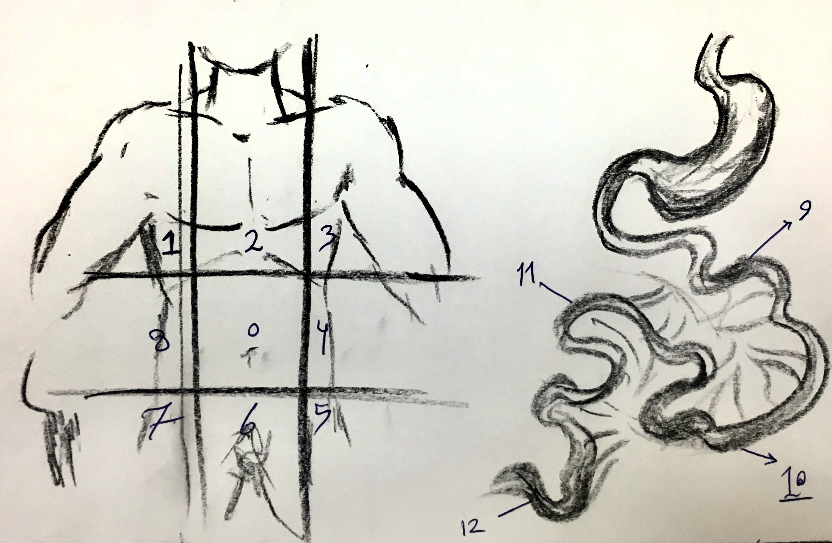
Peritoneal Carcinomatosis Index (PCI) Regions
- CT scan also helps in discovering distant metastatic lessons in the other organs like bone, lungs, and brain.
_Regions.jpg)
References
- ↑ Low RN, Barone RM (2012) Combined diffusion-weighted and gadolinium-enhanced MRI can accurately predict the peritoneal cancer index preoperatively in patients being considered for cytoreductive surgical procedures. Ann Surg Oncol 19 (5):1394-1401. DOI:10.1245/s10434-012-2236-3 PMID: 22302265