Penetrating atherosclerotic aortic ulcer: Difference between revisions
Jump to navigation
Jump to search
(/* 2014 ESC Guidelines on the Diagnosis and Treatment of Aortic Diseases (DO NOT EDIT){{cite journal |vauthors=Erbel R, Aboyans V, Boileau C, Bossone E, Bartolomeo RD, Eggebrecht H, Evangelista A, Falk V, Frank H, Gaemperli O, Grabenwöger M, Haverich...) |
No edit summary |
||
| (65 intermediate revisions by 2 users not shown) | |||
| Line 1: | Line 1: | ||
__NOTOC__ | |||
[[file:Pathogenesis-of-penetrating-atherosclerotic-ulcer-illustration (1).png|200px|right|<ref>Case courtesy of Dr Vincent Tatco, Radiopaedia.org, rID: 48455</ref>]] | |||
{{SI}} | {{SI}} | ||
{{CMG}};'''Associate Editor-In-Chief:''' {{Sahar}} {{CZ}} | |||
{{ | {{SK}}: Penetrating aortic ulcer | ||
==Overview== | ==Overview== | ||
Penetrating atherosclerotic aortic ulcer is ulceration of atheromatous plaque that has eroded the inner, elastic layer of the aortic wall, reached the medial layer, and produced a hematoma in the media. | Penetrating atherosclerotic aortic ulcer is ulceration of [[atheromatous]] [[plaque]] that has eroded the inner, elastic layer of the [[aortic]] wall, reached the medial layer, and produced a [[hematoma]] in the media. | ||
==Historical Perspective== | ==Historical Perspective== | ||
*In 1934, Dr. Shennan was the first to describe the term "penetrating atherosclerotic aortic ulcer".<ref name="Peery1936">{{cite journal|last1=Peery|first1=Thomas M.|title=Dissecting aneurysms of the aorta|journal=American Heart Journal|volume=12|issue=6|year=1936|pages=650–665|issn=00028703|doi=10.1016/S0002-8703(36)91001-3}}</ref> | |||
*In 1986, Dr. Stanson further defined this [[disorder]].<ref name="StansonKazmier1986">{{cite journal|last1=Stanson|first1=Anthony W.|last2=Kazmier|first2=Francis J.|last3=Hollier|first3=Larry H.|last4=Edwards|first4=William D.|last5=Pairolero|first5=Peter C.|last6=Sheedy|first6=Patrick F.|last7=Joyce|first7=John W.|last8=Johnson|first8=Michaël C.|title=Penetrating atherosclerotic ulcers of the thoracic aorta: natural history and clinicopathologic correlations|journal=Annals of Vascular Surgery|volume=1|issue=1|year=1986|pages=15–23|issn=08905096|doi=10.1016/S0890-5096(06)60697-3}}</ref> | |||
==Classification== | ==Classification== | ||
*Penetrating atherosclerotic aortic ulcer is [[Classification|classified]] similarly to the [[aortic dissection]]. | |||
*Learn more about the classification of aortic dissection by clicking '''[[Aortic dissection classification|here]].''' | |||
==Pathophysiology== | ==Pathophysiology== | ||
The | *The [[pathogenesis]] of penetrating atherosclerotic aortic ulcer involves severely diseased [[intima]] in the context of advanced [[atherosclerosis]].<ref name="StansonKazmier1986">{{cite journal|last1=Stanson|first1=Anthony W.|last2=Kazmier|first2=Francis J.|last3=Hollier|first3=Larry H.|last4=Edwards|first4=William D.|last5=Pairolero|first5=Peter C.|last6=Sheedy|first6=Patrick F.|last7=Joyce|first7=John W.|last8=Johnson|first8=Michaël C.|title=Penetrating atherosclerotic ulcers of the thoracic aorta: natural history and clinicopathologic correlations|journal=Annals of Vascular Surgery|volume=1|issue=1|year=1986|pages=15–23|issn=08905096|doi=10.1016/S0890-5096(06)60697-3}}</ref> | ||
*The thickened [[intima]] [[Ulceration|ulcerates]] and this [[ulceration]] propagates through [[intimal]] layer into [[Tunica media|media]] and sometimes it may pass [[Tunica media|media]] through the [[adventitial]] layer of the [[aorta]] and leads to the formation of a periaortic [[pseudoaneurysm]] and even transmural [[aortic rupture]]. | |||
==Causes== | ==Causes== | ||
Penetrating atherosclerotic aortic ulcer may be caused by [ | *Penetrating atherosclerotic aortic ulcer may be caused by advanced [[atherosclerotic disease]].<ref name="StansonKazmier1986">{{cite journal|last1=Stanson|first1=Anthony W.|last2=Kazmier|first2=Francis J.|last3=Hollier|first3=Larry H.|last4=Edwards|first4=William D.|last5=Pairolero|first5=Peter C.|last6=Sheedy|first6=Patrick F.|last7=Joyce|first7=John W.|last8=Johnson|first8=Michaël C.|title=Penetrating atherosclerotic ulcers of the thoracic aorta: natural history and clinicopathologic correlations|journal=Annals of Vascular Surgery|volume=1|issue=1|year=1986|pages=15–23|issn=08905096|doi=10.1016/S0890-5096(06)60697-3}}</ref> | ||
==Differentiating Penetrating | ==Differentiating Penetrating Atherosclerotic Aortic Ulcer from other Diseases== | ||
Penetrating atherosclerotic aortic ulcer must be differentiated from other diseases that cause [ | *Penetrating atherosclerotic aortic ulcer must be [[Differentiate|differentiated]] from other diseases that cause sudden sharp [[chest pain]], [[dyspnea]], and back pain. Such conditions can include [[aortic dissection]], [[Acute coronary syndromes|acute coronary syndrome]], and intramural aortic hematoma. | ||
*Other [[differential diagnosis|differential diagnoses]] of penetrating atherosclerotic aortic ulcer include inflammatory aortic aneurysm and conditions causing inflammatory aortitis such as [[Takayasu arteritis]], [[giant cell arteritis]], [[Behçet disease]], [[Kawasaki disease]], [[rheumatoid arthritis]], [[sarcoidosis]], Cogan syndrome, [[ankylosing spondylitis]], [[systemic lupus erythematosus]], and [[Wegener’s granulomatosis]].<ref name="KotsisSpyropoulos2019">{{cite journal|last1=Kotsis|first1=Thomas|last2=Spyropoulos|first2=Basileios Georgiou|last3=Asaloumidis|first3=Nikolaos|last4=Christoforou|first4=Panagitsa|last5=Katseni|first5=Konstantina|last6=Papaconstantinou|first6=Ioannis|title=Penetrating Atherosclerotic Ulcers of the Abdominal Aorta: A Case Report and Review of the Literature|journal=Vascular Specialist International|volume=35|issue=3|year=2019|pages=152–159|issn=2288-7970|doi=10.5758/vsi.2019.35.3.152}}</ref> | |||
*For more information on the [[differential diagnosis]] of penetrating atherosclerotic aortic ulcer, [[Aortic dissection differential diagnosis|click here]]. | |||
==Epidemiology and Demographics== | ==Epidemiology and Demographics== | ||
The | * The exact [[prevalence]] of penetrating atherosclerotic aortic ulcer is unknown. | ||
*The [[incidence]] of penetrating atherosclerotic aortic ulcer is 1.62 per 100,000 individuals worldwide.<ref name="DeMartinoSen2018">{{cite journal|last1=DeMartino|first1=Randall R.|last2=Sen|first2=Indrani|last3=Huang|first3=Ying|last4=Bower|first4=Thomas C.|last5=Oderich|first5=Gustavo S.|last6=Pochettino|first6=Alberto|last7=Greason|first7=Kevin|last8=Kalra|first8=Manju|last9=Johnstone|first9=Jill|last10=Shuja|first10=Fahad|last11=Harmsen|first11=W. Scott|last12=Macedo|first12=Thanila|last13=Mandrekar|first13=Jay|last14=Chamberlain|first14=Alanna M.|last15=Weiss|first15=Salome|last16=Goodney|first16=Philip P.|last17=Roger|first17=Veronique|title=Population-Based Assessment of the Incidence of Aortic Dissection, Intramural Hematoma, and Penetrating Ulcer, and Its Associated Mortality From 1995 to 2015|journal=Circulation: Cardiovascular Quality and Outcomes|volume=11|issue=8|year=2018|issn=1941-7713|doi=10.1161/CIRCOUTCOMES.118.004689}}</ref> | |||
Unlike typical [[aortic dissection]], penetrating atherosclerotic ulcers most often occur in elderly patients with severe underlying atherosclerosis. | *Unlike typical [[aortic dissection]], penetrating atherosclerotic aortic ulcers most often occur in elderly [[patients]] with severe underlying [[atherosclerosis]].<ref name="KotsisSpyropoulos2019">{{cite journal|last1=Kotsis|first1=Thomas|last2=Spyropoulos|first2=Basileios Georgiou|last3=Asaloumidis|first3=Nikolaos|last4=Christoforou|first4=Panagitsa|last5=Katseni|first5=Konstantina|last6=Papaconstantinou|first6=Ioannis|title=Penetrating Atherosclerotic Ulcers of the Abdominal Aorta: A Case Report and Review of the Literature|journal=Vascular Specialist International|volume=35|issue=3|year=2019|pages=152–159|issn=2288-7970|doi=10.5758/vsi.2019.35.3.152}}</ref> | ||
*It usually affects people in their seventh decade of life or when older. <ref name="StansonKazmier1986">{{cite journal|last1=Stanson|first1=Anthony W.|last2=Kazmier|first2=Francis J.|last3=Hollier|first3=Larry H.|last4=Edwards|first4=William D.|last5=Pairolero|first5=Peter C.|last6=Sheedy|first6=Patrick F.|last7=Joyce|first7=John W.|last8=Johnson|first8=Michaël C.|title=Penetrating atherosclerotic ulcers of the thoracic aorta: natural history and clinicopathologic correlations|journal=Annals of Vascular Surgery|volume=1|issue=1|year=1986|pages=15–23|issn=08905096|doi=10.1016/S0890-5096(06)60697-3}}</ref> | |||
*Penetrating atherosclerotic aortic ulcer affects men at a greater extent than women. | |||
==Risk Factors== | ==Risk Factors== | ||
Common risk factors in the development of penetrating atherosclerotic aortic ulcer include old age, smoking history, male gender, hypertension, coronary artery disease, and | *Common [[Risk factor|risk factors]] in the development of penetrating atherosclerotic aortic ulcer include old [[age]], having a [[smoking]] history, male gender, [[hypertension]], [[Coronary heart disease|coronary artery disease]], and [[hyperlipidemia]].<ref name="StansonKazmier1986">{{cite journal|last1=Stanson|first1=Anthony W.|last2=Kazmier|first2=Francis J.|last3=Hollier|first3=Larry H.|last4=Edwards|first4=William D.|last5=Pairolero|first5=Peter C.|last6=Sheedy|first6=Patrick F.|last7=Joyce|first7=John W.|last8=Johnson|first8=Michaël C.|title=Penetrating atherosclerotic ulcers of the thoracic aorta: natural history and clinicopathologic correlations|journal=Annals of Vascular Surgery|volume=1|issue=1|year=1986|pages=15–23|issn=08905096|doi=10.1016/S0890-5096(06)60697-3}}</ref><ref name="KotsisSpyropoulos2019">{{cite journal|last1=Kotsis|first1=Thomas|last2=Spyropoulos|first2=Basileios Georgiou|last3=Asaloumidis|first3=Nikolaos|last4=Christoforou|first4=Panagitsa|last5=Katseni|first5=Konstantina|last6=Papaconstantinou|first6=Ioannis|title=Penetrating Atherosclerotic Ulcers of the Abdominal Aorta: A Case Report and Review of the Literature|journal=Vascular Specialist International|volume=35|issue=3|year=2019|pages=152–159|issn=2288-7970|doi=10.5758/vsi.2019.35.3.152}}</ref><ref name="CoadyRizzo1999">{{cite journal|last1=Coady|first1=Michael A.|last2=Rizzo|first2=John A.|last3=Elefteriades|first3=John A.|title=PATHOLOGIC VARIANTS OF THORACIC AORTIC DISSECTIONS|journal=Cardiology Clinics|volume=17|issue=4|year=1999|pages=637–657|issn=07338651|doi=10.1016/S0733-8651(05)70106-5}}</ref><ref name="HayashiMatsuoka2000">{{cite journal|last1=Hayashi|first1=Hideyuki|last2=Matsuoka|first2=Yohjiro|last3=Sakamoto|first3=Ichiro|last4=Sueyoshi|first4=Eijun|last5=Okimoto|first5=Tomoaki|last6=Hayashi|first6=Kuniaki|last7=Matsunaga|first7=Naofumi|title=Penetrating Atherosclerotic Ulcer of the Aorta: Imaging Features and Disease Concept|journal=RadioGraphics|volume=20|issue=4|year=2000|pages=995–1005|issn=0271-5333|doi=10.1148/radiographics.20.4.g00jl01995}}</ref> | ||
*Other less common [[risk factors]] include [[bicuspid aortic valve]], prior aortic dilatation, [[connective tissue disorder]], and prior [[aortic]] surgery.<ref name="DeMartinoSen2018">{{cite journal|last1=DeMartino|first1=Randall R.|last2=Sen|first2=Indrani|last3=Huang|first3=Ying|last4=Bower|first4=Thomas C.|last5=Oderich|first5=Gustavo S.|last6=Pochettino|first6=Alberto|last7=Greason|first7=Kevin|last8=Kalra|first8=Manju|last9=Johnstone|first9=Jill|last10=Shuja|first10=Fahad|last11=Harmsen|first11=W. Scott|last12=Macedo|first12=Thanila|last13=Mandrekar|first13=Jay|last14=Chamberlain|first14=Alanna M.|last15=Weiss|first15=Salome|last16=Goodney|first16=Philip P.|last17=Roger|first17=Veronique|title=Population-Based Assessment of the Incidence of Aortic Dissection, Intramural Hematoma, and Penetrating Ulcer, and Its Associated Mortality From 1995 to 2015|journal=Circulation: Cardiovascular Quality and Outcomes|volume=11|issue=8|year=2018|issn=1941-7713|doi=10.1161/CIRCOUTCOMES.118.004689}}</ref> | |||
==Screening== | ==Screening== | ||
There is insufficient evidence to recommend routine screening for penetrating atherosclerotic aortic ulcer. | |||
* There is insufficient evidence to recommend routine [[screening]] for penetrating atherosclerotic aortic ulcer. | |||
==Natural History, Complications, and Prognosis== | ==Natural History, Complications, and Prognosis== | ||
*Penetrating atherosclerotic aortic ulcer starts with the progressive increase in aortic size with subsequent aneurysm formation.<ref name="HayashiMatsuoka2000">{{cite journal|last1=Hayashi|first1=Hideyuki|last2=Matsuoka|first2=Yohjiro|last3=Sakamoto|first3=Ichiro|last4=Sueyoshi|first4=Eijun|last5=Okimoto|first5=Tomoaki|last6=Hayashi|first6=Kuniaki|last7=Matsunaga|first7=Naofumi|title=Penetrating Atherosclerotic Ulcer of the Aorta: Imaging Features and Disease Concept|journal=RadioGraphics|volume=20|issue=4|year=2000|pages=995–1005|issn=0271-5333|doi=10.1148/radiographics.20.4.g00jl01995}}</ref><ref name="NathanBoonn2012">{{cite journal|last1=Nathan|first1=Derek P.|last2=Boonn|first2=William|last3=Lai|first3=Eric|last4=Wang|first4=Grace J.|last5=Desai|first5=Nimesh|last6=Woo|first6=Edward Y.|last7=Fairman|first7=Ronald M.|last8=Jackson|first8=Benjamin M.|title=Presentation, complications, and natural history of penetrating atherosclerotic ulcer disease|journal=Journal of Vascular Surgery|volume=55|issue=1|year=2012|pages=10–15|issn=07415214|doi=10.1016/j.jvs.2011.08.005}}</ref> | *The natural history of penetrating atherosclerotic aortic ulcer include an elderly with multiple [[risk factors]] for advanced [[atherosclerosis]] who presents with acute onset [[chest pain]] and [[dyspnea]] which may progress and lead to [[aortic dissection]].<ref name="UedaChin2012">{{cite journal|last1=Ueda|first1=Takuya|last2=Chin|first2=Anne|last3=Petrovitch|first3=Ivan|last4=Fleischmann|first4=Dominik|title=A pictorial review of acute aortic syndrome: discriminating and overlapping features as revealed by ECG-gated multidetector-row CT angiography|journal=Insights into Imaging|volume=3|issue=6|year=2012|pages=561–571|issn=1869-4101|doi=10.1007/s13244-012-0195-7}}</ref> | ||
*If left untreated, patients with penetrating atherosclerotic aortic ulcer may progress to develop an intramural hematoma, pseudoaneurysm, or even aortic rupture | *Penetrating atherosclerotic aortic ulcer starts with the progressive increase in aortic size with subsequent [[aneurysm]] formation.<ref name="HayashiMatsuoka2000">{{cite journal|last1=Hayashi|first1=Hideyuki|last2=Matsuoka|first2=Yohjiro|last3=Sakamoto|first3=Ichiro|last4=Sueyoshi|first4=Eijun|last5=Okimoto|first5=Tomoaki|last6=Hayashi|first6=Kuniaki|last7=Matsunaga|first7=Naofumi|title=Penetrating Atherosclerotic Ulcer of the Aorta: Imaging Features and Disease Concept|journal=RadioGraphics|volume=20|issue=4|year=2000|pages=995–1005|issn=0271-5333|doi=10.1148/radiographics.20.4.g00jl01995}}</ref><ref name="NathanBoonn2012">{{cite journal|last1=Nathan|first1=Derek P.|last2=Boonn|first2=William|last3=Lai|first3=Eric|last4=Wang|first4=Grace J.|last5=Desai|first5=Nimesh|last6=Woo|first6=Edward Y.|last7=Fairman|first7=Ronald M.|last8=Jackson|first8=Benjamin M.|title=Presentation, complications, and natural history of penetrating atherosclerotic ulcer disease|journal=Journal of Vascular Surgery|volume=55|issue=1|year=2012|pages=10–15|issn=07415214|doi=10.1016/j.jvs.2011.08.005}}</ref> | ||
*Prognosis is generally poor and even worse than that of aortic dissection. | *If left untreated, [[patients]] with penetrating atherosclerotic aortic ulcer may progress to develop an intramural hematoma, [[pseudoaneurysm]], acute [[aortic dissection]], or even [[aortic rupture]]. | ||
*[[Prognosis]] is generally poor and even worse than that of [[aortic dissection]]. | |||
==Diagnosis== | ==Diagnosis== | ||
===Diagnostic Study of Choice=== | ===Diagnostic Study of Choice=== | ||
*Table below provides a comparison of diagnostic imaging studies for the diagnosis of penetrating atherosclerotic aortic ulcer:<ref name="pmid25173340">{{cite journal |vauthors=Erbel R, Aboyans V, Boileau C, Bossone E, Bartolomeo RD, Eggebrecht H, Evangelista A, Falk V, Frank H, Gaemperli O, Grabenwöger M, Haverich A, Iung B, Manolis AJ, Meijboom F, Nienaber CA, Roffi M, Rousseau H, Sechtem U, Sirnes PA, Allmen RS, Vrints CJ |title=2014 ESC Guidelines on the diagnosis and treatment of aortic diseases: Document covering acute and chronic aortic diseases of the thoracic and abdominal aorta of the adult. The Task Force for the Diagnosis and Treatment of Aortic Diseases of the European Society of Cardiology (ESC) |journal=Eur. Heart J. |volume=35 |issue=41 |pages=2873–926 |date=November 2014 |pmid=25173340 |doi=10.1093/eurheartj/ehu281 |url=}}</ref> | |||
* Contrast-enhanced [[CT|CT imaging]] is the [[diagnostic study of choice]] for [[diagnosis]] of penetrating atherosclerotic aortic ulcer. | |||
*Table below provides a comparison of [[diagnostic]] imaging studies for the [[diagnosis]] of penetrating atherosclerotic aortic ulcer:<ref name="pmid25173340">{{cite journal |vauthors=Erbel R, Aboyans V, Boileau C, Bossone E, Bartolomeo RD, Eggebrecht H, Evangelista A, Falk V, Frank H, Gaemperli O, Grabenwöger M, Haverich A, Iung B, Manolis AJ, Meijboom F, Nienaber CA, Roffi M, Rousseau H, Sechtem U, Sirnes PA, Allmen RS, Vrints CJ |title=2014 ESC Guidelines on the diagnosis and treatment of aortic diseases: Document covering acute and chronic aortic diseases of the thoracic and abdominal aorta of the adult. The Task Force for the Diagnosis and Treatment of Aortic Diseases of the European Society of Cardiology (ESC) |journal=Eur. Heart J. |volume=35 |issue=41 |pages=2873–926 |date=November 2014 |pmid=25173340 |doi=10.1093/eurheartj/ehu281 |url=}}</ref> | |||
{| class="wikitable" | {| class="wikitable" | ||
|+ | |+ | ||
|- | |- | ||
| align="center" style="background:#4479BA; color: #FFFFFF;" |'''Diagnostic Modality''' | | align="center" style="background:#4479BA; color: #FFFFFF;" |'''Diagnostic Modality''' | ||
! align="center" style="background:#4479BA; color: #FFFFFF;" |Diagnostic Value | |||
|- | |- | ||
| | | | ||
*[[Transthoracic echocardiography|Transthoracic Echocardiography]] | *[[Transthoracic echocardiography|Transthoracic Echocardiography]] | ||
| Line 45: | Line 60: | ||
|- | |- | ||
| | | | ||
* Transesophageal echocardiography | *[[Transesophageal echocardiography (TEE)|Transesophageal echocardiography]] | ||
| | | | ||
* Moderate | * Moderate | ||
|- | |- | ||
| | | | ||
* CT Scan | *[[CT Scan]] | ||
| | | | ||
* Excellent | * Excellent | ||
|- | |- | ||
| | | | ||
* MRI | *[[MRI]] | ||
| | | | ||
* Excellent | * Excellent | ||
|} | |} | ||
* | |||
* | |||
===History and Symptoms=== | ===History and Symptoms=== | ||
*History and [[symptoms]] of pentrating atherosclerotic aortic ulcer may greatly overlap with those of [[aortic dissection]]. Nevertheless, [[lesion]] is usually localized and does not result in radiating [[pain]]. [[Patients]] may also remain asymptomatic. The most common [[symptoms]] include [[chest pain]] and [[back pain]].<ref name="BossoneLaBounty2018">{{cite journal|last1=Bossone|first1=Eduardo|last2=LaBounty|first2=Troy M|last3=Eagle|first3=Kim A|title=Acute aortic syndromes: diagnosis and management, an update|journal=European Heart Journal|volume=39|issue=9|year=2018|pages=739–749d|issn=0195-668X|doi=10.1093/eurheartj/ehx319}}</ref> | |||
===Physical Examination=== | ===Physical Examination=== | ||
*Physical examination of [[patients]] with penetrating atherosclerotic aortic ulcer may overlap with that of [[aortic dissection]]. Nevertheless, due to the localized nature of the [[lesion]], [[signs]], such as absent pulse or [[Diastolic murmurs|diastolic murmur]] of [[aortic regurgitation]] are unlikely. Possible physical examination findings include [[hypotension]] and shock especially in the case of accompanying [[aortic rupture]].<ref name="BossoneLaBounty2018">{{cite journal|last1=Bossone|first1=Eduardo|last2=LaBounty|first2=Troy M|last3=Eagle|first3=Kim A|title=Acute aortic syndromes: diagnosis and management, an update|journal=European Heart Journal|volume=39|issue=9|year=2018|pages=739–749d|issn=0195-668X|doi=10.1093/eurheartj/ehx319}}</ref> | |||
===Laboratory Findings=== | ===Laboratory Findings=== | ||
== | *There are no [[diagnostic]] laboratory findings associated with penetrating atherosclerotic aortic ulcer. | ||
===Electrocardiogram=== | |||
*There are no [[ECG]] findings associated with penetrating atherosclerotic aortic ulcer. | |||
{| align="right" | |||
|[[File:Penetrating-aortic-atherosclerotic-ulcer-with-false-aneurysm.jpg|thumb|none|200px|Penetrating atherosclerotic aortic ulcer<ref>Case courtesy of A.Prof Frank Gaillard, Radiopaedia.org, rID: 10163</ref>]] | |||
|} | |||
===X-ray=== | |||
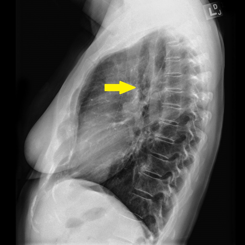
*Findings associated with penetrating atherosclerotic aortic ulcer on an [[x-ray]] may include the widening of the thoracoabdominal aortic silhouette, diffuse or focal enlargement of [[thoracic]] [[descending aorta]], [[pleural effusion]], and deviated [[trachea]].<ref name="EggebrechtBaumgart2003">{{cite journal|last1=Eggebrecht|first1=Holger|last2=Baumgart|first2=Dietrich|last3=Schmermund|first3=Axel|last4=Herold|first4=Ulf|last5=Hunold|first5=Peter|last6=Jakob|first6=Heinz|last7=Erbel|first7=Raimund|title=Penetrating atherosclerotic ulcer of the aorta: treatment by endovascular stent-graft placement|journal=Current Opinion in Cardiology|volume=18|issue=6|year=2003|pages=431–435|issn=0268-4705|doi=10.1097/00001573-200311000-00002}}</ref> | |||
*A normal [[chest]] [[x-ray]] does not rule out the [[diagnosis]] of penetrating atherosclerotic aortic ulcer.<ref name="KyawSadiq2016">{{cite journal|last1=Kyaw|first1=Htoo|last2=Sadiq|first2=Sanah|last3=Chowdhury|first3=Arnab|last4=Gholamrezaee|first4=Rashin|last5=Yoe|first5=Linus|title=An uncommon cause of chest pain – penetrating atherosclerotic aortic ulcer|journal=Journal of Community Hospital Internal Medicine Perspectives|volume=6|issue=3|year=2016|pages=31506|issn=2000-9666|doi=10.3402/jchimp.v6.31506}}</ref> | |||
===Echocardigraphy=== | ===Echocardigraphy=== | ||
* | *Tans-esophageal [[echocardiography]] has been approved to be sensitive and specific for the [[diagnosis]] of [[aortic]] diseases.<ref name="pmid8668776">{{cite journal |vauthors=Sommer T, Fehske W, Holzknecht N, Smekal AV, Keller E, Lutterbey G, Kreft B, Kuhl C, Gieseke J, Abu-Ramadan D, Schild H |title=Aortic dissection: a comparative study of diagnosis with spiral CT, multiplanar transesophageal echocardiography, and MR imaging |journal=Radiology |volume=199 |issue=2 |pages=347–52 |date=May 1996 |pmid=8668776 |doi=10.1148/radiology.199.2.8668776 |url=}}</ref> | ||
===CT Scan=== | ===CT Scan=== | ||
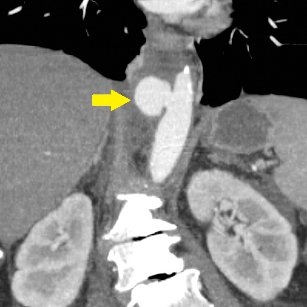
*CT scan imaging with intravenous contrast is the diagnostic study of choice for diagnosis of penetrating atherosclerotic aortic ulcer.<ref name="HayashiMatsuoka2000">{{cite journal|last1=Hayashi|first1=Hideyuki|last2=Matsuoka|first2=Yohjiro|last3=Sakamoto|first3=Ichiro|last4=Sueyoshi|first4=Eijun|last5=Okimoto|first5=Tomoaki|last6=Hayashi|first6=Kuniaki|last7=Matsunaga|first7=Naofumi|title=Penetrating Atherosclerotic Ulcer of the Aorta: Imaging Features and Disease Concept|journal=RadioGraphics|volume=20|issue=4|year=2000|pages=995–1005|issn=0271-5333|doi=10.1148/radiographics.20.4.g00jl01995}}</ref> | *[[CT scan|CT scan imaging]] with intravenous contrast is the [[Diagnosis|diagnostic]] study of choice for [[diagnosis]] of penetrating atherosclerotic aortic ulcer.<ref name="HayashiMatsuoka2000">{{cite journal|last1=Hayashi|first1=Hideyuki|last2=Matsuoka|first2=Yohjiro|last3=Sakamoto|first3=Ichiro|last4=Sueyoshi|first4=Eijun|last5=Okimoto|first5=Tomoaki|last6=Hayashi|first6=Kuniaki|last7=Matsunaga|first7=Naofumi|title=Penetrating Atherosclerotic Ulcer of the Aorta: Imaging Features and Disease Concept|journal=RadioGraphics|volume=20|issue=4|year=2000|pages=995–1005|issn=0271-5333|doi=10.1148/radiographics.20.4.g00jl01995}}</ref> | ||
*Findings suggestive of penetrating atherosclerotic aortic ulcer include a localized ulcer passing from intima into aortic wall or contrast leak through a calcified plaque. | *Findings suggestive of penetrating atherosclerotic aortic ulcer include a localized [[ulcer]] passing from [[intima]] into aortic wall or contrast leak through a calcified plaque. | ||
*It usually affects mid to distal third of descending aorta. | *It usually affects mid to distal third of [[descending aorta]]. | ||
*Ulcer is usually defined by focal thickening or enhancement of aortic wall. | *Ulcer is usually defined by focal thickening or enhancement of the aortic wall. | ||
{| align="right" | |||
|[[File:Penetrating-aortic-atherosclerotic-ulcer-with-false-aneurysm (2).jpg|thumb|none|200px|contrast-enhanced CT<ref>Case courtesy of A.Prof Frank Gaillard, Radiopaedia.org, rID: 10163</ref>]] | |||
|} | |||
===MRI=== | ===MRI=== | ||
*MRI study is superior to conventional CT scan in the | *[[MRI]] study is superior to conventional [[CT scan]] in the [[diagnosis]] of penetrating atherosclerotic aortic ulcer.<ref name="HarrisBis1994">{{cite journal|last1=Harris|first1=James A.|last2=Bis|first2=Kostaki G.|last3=Glover|first3=John L.|last4=Bendick|first4=Phillip J.|last5=Shetty|first5=Anil|last6=Brown|first6=O.William|title=Penetrating atherosclerotic ulcers of the aorta|journal=Journal of Vascular Surgery|volume=19|issue=1|year=1994|pages=90–99|issn=07415214|doi=10.1016/S0741-5214(94)70124-5}}</ref> | ||
*Finding suggestive of the diagnosis includes a well-defined ulcer with flow void phenomenon on T1 images. | *Finding suggestive of the [[diagnosis]] includes a well-defined ulcer with flow void phenomenon on T1 images. | ||
===Other Imaging Findings=== | |||
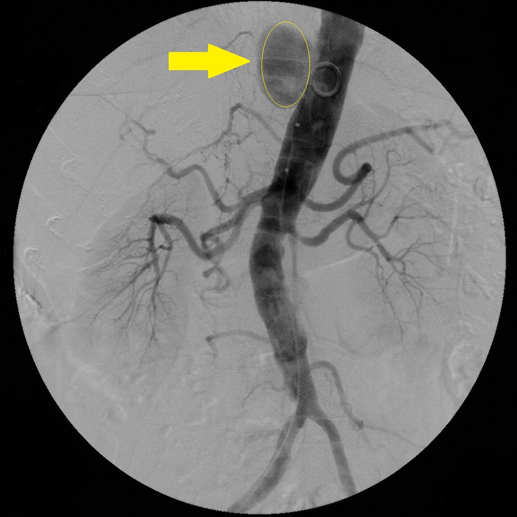
*Findings associated with penetrating atherosclerotic aortic ulcer on a [[CT angiography]] may include the presence of false [[aneurysm]]. | |||
===Other Diagnostic Studies=== | |||
*There are no other [[diagnostic]] studies associated with penetrating atherosclerotic aortic ulcer. | |||
==Treatment== | ==Treatment== | ||
{| align="right" | |||
|[[File:Penetrating-aortic-atherosclerotic-ulcer-with-false-aneurysm (1).jpg|thumb|none|200px|Penetrating aortic atherosclerotic ulcer with false aneurysm<ref>Case courtesy of A.Prof Frank Gaillard, Radiopaedia.org, rID: 10163</ref>]] | |||
|} | |||
===Medical Therapy=== | ===Medical Therapy=== | ||
*The treatment aims at preventing the penetrating atherosclerotic aortic ulcer to progress into [[aortic dissection]]. [[Indication (medicine)|Indications]] for treatment interventions include recurrent and refractory [[pain]], [[signs]] of contained rupture, rapidly growing aortic ulcer, and presence of periaortic hematoma or [[pleural effusion]].<ref name="GanahaMiller2002">{{cite journal|last1=Ganaha|first1=Fumikiyo|last2=Miller|first2=D. Craig|last3=Sugimoto|first3=Koji|last4=Do|first4=Young Soo|last5=Minamiguchi|first5=Hiroki|last6=Saito|first6=Haruo|last7=Mitchell|first7=R. Scott|last8=Dake|first8=Michael D.|title=Prognosis of Aortic Intramural Hematoma With and Without Penetrating Atherosclerotic Ulcer|journal=Circulation|volume=106|issue=3|year=2002|pages=342–348|issn=0009-7322|doi=10.1161/01.CIR.0000022164.26075.5A}}</ref><ref name="EggebrechtHerold2006">{{cite journal|last1=Eggebrecht|first1=Holger|last2=Herold|first2=Ulf|last3=Schmermund|first3=Axel|last4=Lind|first4=Alexander Y.|last5=Kuhnt|first5=Oliver|last6=Martini|first6=Stefan|last7=Kühl|first7=Hilmar|last8=Kienbaum|first8=Peter|last9=Peters|first9=Jürgen|last10=Jakob|first10=Heinz|last11=Erbel|first11=Raimund|last12=Baumgart|first12=Dietrich|title=Endovascular stent-graft treatment of penetrating aortic ulcer|journal=American Heart Journal|volume=151|issue=2|year=2006|pages=530–536|issn=00028703|doi=10.1016/j.ahj.2005.05.020}}</ref> | |||
*Early interventions are indicated in ulcer with a diameter greater than 20 mm. | |||
===Surgery=== | ===Surgery=== | ||
*[[Surgical]] interventions may be [[Indication (medicine)|indicated]] in penetrating atherosclerotic aortic ulcer depending on the anatomic location of the ulcer, clinical presentation and [[comorbidities]]. However, since most of the [[patients]] are poor [[surgical]] candidates due to advanced age and associated [[comorbidities]], thoracic endovascular aortic repair (TEVAR) is used more frequently.<ref name="EggebrechtHerold2006">{{cite journal|last1=Eggebrecht|first1=Holger|last2=Herold|first2=Ulf|last3=Schmermund|first3=Axel|last4=Lind|first4=Alexander Y.|last5=Kuhnt|first5=Oliver|last6=Martini|first6=Stefan|last7=Kühl|first7=Hilmar|last8=Kienbaum|first8=Peter|last9=Peters|first9=Jürgen|last10=Jakob|first10=Heinz|last11=Erbel|first11=Raimund|last12=Baumgart|first12=Dietrich|title=Endovascular stent-graft treatment of penetrating aortic ulcer|journal=American Heart Journal|volume=151|issue=2|year=2006|pages=530–536|issn=00028703|doi=10.1016/j.ahj.2005.05.020}}</ref><ref name="DemersMiller2004">{{cite journal|last1=Demers|first1=Philippe|last2=Miller|first2=D.Craig|last3=Mitchell|first3=R.Scott|last4=Kee|first4=Stephen T|last5=Chagonjian|first5=Lynn|last6=Dake|first6=Michael D|title=Stent-graft repair of penetrating atherosclerotic ulcers in the descending thoracic aorta: mid-term results|journal=The Annals of Thoracic Surgery|volume=77|issue=1|year=2004|pages=81–86|issn=00034975|doi=10.1016/S0003-4975(03)00816-6}}</ref> | |||
===Primary Prevention=== | ===Primary Prevention=== | ||
There are no established measures for the primary prevention of penetrating atherosclerotic aortic ulcer. | *There are no established measures for the primary prevention of penetrating atherosclerotic aortic ulcer. | ||
===Secondary Prevention=== | ===Secondary Prevention=== | ||
There are no established measures for the secondary prevention of penetrating atherosclerotic aortic ulcer. | *There are no established measures for the secondary prevention of penetrating atherosclerotic aortic ulcer. | ||
== | ==Guidelines== | ||
===2014 ESC Guidelines on the Diagnosis and Treatment of Aortic Diseases (DO NOT EDIT)<ref name="pmid25173340">{{cite journal |vauthors=Erbel R, Aboyans V, Boileau C, Bossone E, Bartolomeo RD, Eggebrecht H, Evangelista A, Falk V, Frank H, Gaemperli O, Grabenwöger M, Haverich A, Iung B, Manolis AJ, Meijboom F, Nienaber CA, Roffi M, Rousseau H, Sechtem U, Sirnes PA, Allmen RS, Vrints CJ |title=2014 ESC Guidelines on the diagnosis and treatment of aortic diseases: Document covering acute and chronic aortic diseases of the thoracic and abdominal aorta of the adult. The Task Force for the Diagnosis and Treatment of Aortic Diseases of the European Society of Cardiology (ESC) |journal=Eur. Heart J. |volume=35 |issue=41 |pages=2873–926 |date=November 2014 |pmid=25173340 |doi=10.1093/eurheartj/ehu281 |url=}}</ref>=== | ===2014 ESC Guidelines on the Diagnosis and Treatment of Aortic Diseases (DO NOT EDIT)<ref name="pmid25173340">{{cite journal |vauthors=Erbel R, Aboyans V, Boileau C, Bossone E, Bartolomeo RD, Eggebrecht H, Evangelista A, Falk V, Frank H, Gaemperli O, Grabenwöger M, Haverich A, Iung B, Manolis AJ, Meijboom F, Nienaber CA, Roffi M, Rousseau H, Sechtem U, Sirnes PA, Allmen RS, Vrints CJ |title=2014 ESC Guidelines on the diagnosis and treatment of aortic diseases: Document covering acute and chronic aortic diseases of the thoracic and abdominal aorta of the adult. The Task Force for the Diagnosis and Treatment of Aortic Diseases of the European Society of Cardiology (ESC) |journal=Eur. Heart J. |volume=35 |issue=41 |pages=2873–926 |date=November 2014 |pmid=25173340 |doi=10.1093/eurheartj/ehu281 |url=}}</ref>=== | ||
{| border="3" | {| border="3" | ||
|+ | |+ | ||
! style="background: #FFFF00; width: 150px;" | | ! style="background: #FFFF00; width: 150px;" | Recommendations !! style="background: #FFFF00; width: 150px;" | Class !! style="background: #FFFF00; width: 150px;" | Level | ||
|- | |- | ||
! style="padding: 5px 5px; background: #FFFFE0; " align="left" |<nowiki>"</nowiki>In all patients with PAU, medical therapy including pain relief and blood pressure control is recommended.<nowiki>"</nowiki> | ! style="padding: 5px 5px; background: #FFFFE0; " align="left" | | ||
* <nowiki>"</nowiki>In all [[patients]] with PAU, medical therapy including pain relief and [[blood pressure]] control is recommended.<nowiki>"</nowiki> | |||
| style="padding: 5px 5px; background: #90EE90;" align="center" |'''I''' | | style="padding: 5px 5px; background: #90EE90;" align="center" |'''I''' | ||
| style="padding: 5px 5px; background: #6495ED;" align="center"|{{fontcolor|#FFF|C}} | | style="padding: 5px 5px; background: #6495ED;" align="center"|{{fontcolor|#FFF|C}} | ||
|- | |- | ||
! style="padding: 5px 5px; background: " align="left" |<nowiki>"</nowiki>In the case of Type A PAU, surgery should be considered.<nowiki>"</nowiki> | ! style="padding: 5px 5px; background: " align="left" | | ||
* <nowiki>"</nowiki>In the case of Type A PAU, [[surgery]] should be considered.<nowiki>"</nowiki> | |||
| style="padding: 5px 5px; background: #FFA500;" align="center" |'''IIa''' | | style="padding: 5px 5px; background: #FFA500;" align="center" |'''IIa''' | ||
| style="padding: 5px 5px; background: #6495ED;" align="center" |{{fontcolor|#FFF|C}} | | style="padding: 5px 5px; background: #6495ED;" align="center" |{{fontcolor|#FFF|C}} | ||
|- | |- | ||
! style="padding: 5px 5px; background: #FFFFE0; " align="left" |<nowiki>"</nowiki>In the case of Type B PAU, initial medical therapy under careful surveillance is recommended.<nowiki>"</nowiki> | ! style="padding: 5px 5px; background: #FFFFE0; " align="left" | | ||
* <nowiki>"</nowiki>In the case of Type B PAU, initial medical therapy under careful surveillance is recommended.<nowiki>"</nowiki> | |||
| style="padding: 5px 5px; background: #90EE90;" align="center" |'''I''' | | style="padding: 5px 5px; background: #90EE90;" align="center" |'''I''' | ||
| style="padding: 5px 5px; background: #6495ED;" align="center" |{{fontcolor|#FFF|C}} | | style="padding: 5px 5px; background: #6495ED;" align="center" |{{fontcolor|#FFF|C}} | ||
|- | |- | ||
! style="padding: 5px 5px; background: " align="left" |<nowiki>"</nowiki>In uncomplicated Type B PAU, repetitive imaging (MRI or CT) is indicated.<nowiki>"</nowiki> | ! style="padding: 5px 5px; background: " align="left" | | ||
* <nowiki>"</nowiki>In uncomplicated Type B PAU, repetitive [[imaging]] ([[MRI]] or [[CT]]) is [[Indication (medicine)|indicated]].<nowiki>"</nowiki> | |||
| style="padding: 5px 5px; background: #90EE90;" align="center" |'''I''' | | style="padding: 5px 5px; background: #90EE90;" align="center" |'''I''' | ||
| style="padding: 5px 5px; background: #6495ED;" align="center" |{{fontcolor|#FFF|C}} | | style="padding: 5px 5px; background: #6495ED;" align="center" |{{fontcolor|#FFF|C}} | ||
|- | |- | ||
! style="padding: 5px 5px; background: #FFFFE0;" align="left" |<nowiki>"</nowiki>In complicated Type B PAU, TEVAR should be considered.<nowiki>"</nowiki> | ! style="padding: 5px 5px; background: #FFFFE0;" align="left" | | ||
* <nowiki>"</nowiki>In complicated Type B PAU, TEVAR should be considered.<nowiki>"</nowiki> | |||
| style="padding: 5px 5px; background: #FFA500;" align="center" |'''IIa''' | | style="padding: 5px 5px; background: #FFA500;" align="center" |'''IIa''' | ||
| style="padding: 5px 5px; background: #6495ED;" align="center" |{{fontcolor|#FFF|C}} | | style="padding: 5px 5px; background: #6495ED;" align="center" |{{fontcolor|#FFF|C}} | ||
|- | |- | ||
! style="padding: 5px 5px;" align="left" |<nowiki>"</nowiki>In complicated Type B PAU, surgery may be considered.<nowiki>"</nowiki> | ! style="padding: 5px 5px;" align="left" | | ||
* <nowiki>"</nowiki>In complicated Type B PAU, [[surgery]] may be considered.<nowiki>"</nowiki> | |||
| style="padding: 5px 5px; background: #FFA500;" align="center" |'''IIb''' | | style="padding: 5px 5px; background: #FFA500;" align="center" |'''IIb''' | ||
| style="padding: 5px 5px; background: #6495ED;" align="center" |{{fontcolor|#FFF|C}} | | style="padding: 5px 5px; background: #6495ED;" align="center" |{{fontcolor|#FFF|C}} | ||
| Line 115: | Line 164: | ||
<small><small> | <small><small> | ||
'''Abbreviations:''' '''CT''': computed tomography; '''MRI''': magnetic resonance imaging; '''PAU''': penetrating aortic ulcer; '''TEVAR''': thoracic endovascular aortic repair. | '''Abbreviations:''' '''CT''': computed tomography; '''MRI''': magnetic resonance imaging; '''PAU''': penetrating aortic ulcer; '''TEVAR''': thoracic endovascular aortic repair. | ||
<small><small> | </small></small> | ||
==See Also== | ==See Also== | ||
*[[Acute aortic syndrome]] | |||
*[[Aortic dissection]] | *[[Aortic dissection]] | ||
*[[Aortic intramural hematoma]] | *[[Aortic intramural hematoma]] | ||
*[[Aortic rupture]] | |||
==External Links== | ==External Links== | ||
*[http://goldminer.arrs.org/search.php?query=Penetrating%20atherosclerotic%20aortic%20ulcer Goldminer: Penetrating atherosclerotic aortic ulcer] | *[http://goldminer.arrs.org/search.php?query=Penetrating%20atherosclerotic%20aortic%20ulcer Goldminer: Penetrating atherosclerotic aortic ulcer] | ||
Latest revision as of 04:41, 23 September 2020
![[1]](/images/0/08/Pathogenesis-of-penetrating-atherosclerotic-ulcer-illustration_%281%29.png)
Editor-In-Chief: C. Michael Gibson, M.S., M.D. [1];Associate Editor-In-Chief: Sahar Memar Montazerin, M.D.[2] Cafer Zorkun, M.D., Ph.D. [3]
Synonyms and keywords:: Penetrating aortic ulcer
Overview
Penetrating atherosclerotic aortic ulcer is ulceration of atheromatous plaque that has eroded the inner, elastic layer of the aortic wall, reached the medial layer, and produced a hematoma in the media.
Historical Perspective
- In 1934, Dr. Shennan was the first to describe the term "penetrating atherosclerotic aortic ulcer".[2]
- In 1986, Dr. Stanson further defined this disorder.[3]
Classification
- Penetrating atherosclerotic aortic ulcer is classified similarly to the aortic dissection.
- Learn more about the classification of aortic dissection by clicking here.
Pathophysiology
- The pathogenesis of penetrating atherosclerotic aortic ulcer involves severely diseased intima in the context of advanced atherosclerosis.[3]
- The thickened intima ulcerates and this ulceration propagates through intimal layer into media and sometimes it may pass media through the adventitial layer of the aorta and leads to the formation of a periaortic pseudoaneurysm and even transmural aortic rupture.
Causes
- Penetrating atherosclerotic aortic ulcer may be caused by advanced atherosclerotic disease.[3]
Differentiating Penetrating Atherosclerotic Aortic Ulcer from other Diseases
- Penetrating atherosclerotic aortic ulcer must be differentiated from other diseases that cause sudden sharp chest pain, dyspnea, and back pain. Such conditions can include aortic dissection, acute coronary syndrome, and intramural aortic hematoma.
- Other differential diagnoses of penetrating atherosclerotic aortic ulcer include inflammatory aortic aneurysm and conditions causing inflammatory aortitis such as Takayasu arteritis, giant cell arteritis, Behçet disease, Kawasaki disease, rheumatoid arthritis, sarcoidosis, Cogan syndrome, ankylosing spondylitis, systemic lupus erythematosus, and Wegener’s granulomatosis.[4]
- For more information on the differential diagnosis of penetrating atherosclerotic aortic ulcer, click here.
Epidemiology and Demographics
- The exact prevalence of penetrating atherosclerotic aortic ulcer is unknown.
- The incidence of penetrating atherosclerotic aortic ulcer is 1.62 per 100,000 individuals worldwide.[5]
- Unlike typical aortic dissection, penetrating atherosclerotic aortic ulcers most often occur in elderly patients with severe underlying atherosclerosis.[4]
- It usually affects people in their seventh decade of life or when older. [3]
- Penetrating atherosclerotic aortic ulcer affects men at a greater extent than women.
Risk Factors
- Common risk factors in the development of penetrating atherosclerotic aortic ulcer include old age, having a smoking history, male gender, hypertension, coronary artery disease, and hyperlipidemia.[3][4][6][7]
- Other less common risk factors include bicuspid aortic valve, prior aortic dilatation, connective tissue disorder, and prior aortic surgery.[5]
Screening
- There is insufficient evidence to recommend routine screening for penetrating atherosclerotic aortic ulcer.
Natural History, Complications, and Prognosis
- The natural history of penetrating atherosclerotic aortic ulcer include an elderly with multiple risk factors for advanced atherosclerosis who presents with acute onset chest pain and dyspnea which may progress and lead to aortic dissection.[8]
- Penetrating atherosclerotic aortic ulcer starts with the progressive increase in aortic size with subsequent aneurysm formation.[7][9]
- If left untreated, patients with penetrating atherosclerotic aortic ulcer may progress to develop an intramural hematoma, pseudoaneurysm, acute aortic dissection, or even aortic rupture.
- Prognosis is generally poor and even worse than that of aortic dissection.
Diagnosis
Diagnostic Study of Choice
- Contrast-enhanced CT imaging is the diagnostic study of choice for diagnosis of penetrating atherosclerotic aortic ulcer.
- Table below provides a comparison of diagnostic imaging studies for the diagnosis of penetrating atherosclerotic aortic ulcer:[10]
| Diagnostic Modality | Diagnostic Value |
|---|---|
| |
| |
| |
|
History and Symptoms
- History and symptoms of pentrating atherosclerotic aortic ulcer may greatly overlap with those of aortic dissection. Nevertheless, lesion is usually localized and does not result in radiating pain. Patients may also remain asymptomatic. The most common symptoms include chest pain and back pain.[11]
Physical Examination
- Physical examination of patients with penetrating atherosclerotic aortic ulcer may overlap with that of aortic dissection. Nevertheless, due to the localized nature of the lesion, signs, such as absent pulse or diastolic murmur of aortic regurgitation are unlikely. Possible physical examination findings include hypotension and shock especially in the case of accompanying aortic rupture.[11]
Laboratory Findings
- There are no diagnostic laboratory findings associated with penetrating atherosclerotic aortic ulcer.
Electrocardiogram
- There are no ECG findings associated with penetrating atherosclerotic aortic ulcer.
 |
X-ray
- Findings associated with penetrating atherosclerotic aortic ulcer on an x-ray may include the widening of the thoracoabdominal aortic silhouette, diffuse or focal enlargement of thoracic descending aorta, pleural effusion, and deviated trachea.[13]
- A normal chest x-ray does not rule out the diagnosis of penetrating atherosclerotic aortic ulcer.[14]
Echocardigraphy
- Tans-esophageal echocardiography has been approved to be sensitive and specific for the diagnosis of aortic diseases.[15]
CT Scan
- CT scan imaging with intravenous contrast is the diagnostic study of choice for diagnosis of penetrating atherosclerotic aortic ulcer.[7]
- Findings suggestive of penetrating atherosclerotic aortic ulcer include a localized ulcer passing from intima into aortic wall or contrast leak through a calcified plaque.
- It usually affects mid to distal third of descending aorta.
- Ulcer is usually defined by focal thickening or enhancement of the aortic wall.
 |
MRI
- MRI study is superior to conventional CT scan in the diagnosis of penetrating atherosclerotic aortic ulcer.[17]
- Finding suggestive of the diagnosis includes a well-defined ulcer with flow void phenomenon on T1 images.
Other Imaging Findings
- Findings associated with penetrating atherosclerotic aortic ulcer on a CT angiography may include the presence of false aneurysm.
Other Diagnostic Studies
- There are no other diagnostic studies associated with penetrating atherosclerotic aortic ulcer.
Treatment
 |
Medical Therapy
- The treatment aims at preventing the penetrating atherosclerotic aortic ulcer to progress into aortic dissection. Indications for treatment interventions include recurrent and refractory pain, signs of contained rupture, rapidly growing aortic ulcer, and presence of periaortic hematoma or pleural effusion.[19][20]
- Early interventions are indicated in ulcer with a diameter greater than 20 mm.
Surgery
- Surgical interventions may be indicated in penetrating atherosclerotic aortic ulcer depending on the anatomic location of the ulcer, clinical presentation and comorbidities. However, since most of the patients are poor surgical candidates due to advanced age and associated comorbidities, thoracic endovascular aortic repair (TEVAR) is used more frequently.[20][21]
Primary Prevention
- There are no established measures for the primary prevention of penetrating atherosclerotic aortic ulcer.
Secondary Prevention
- There are no established measures for the secondary prevention of penetrating atherosclerotic aortic ulcer.
Guidelines
2014 ESC Guidelines on the Diagnosis and Treatment of Aortic Diseases (DO NOT EDIT)[10]
| Recommendations | Class | Level |
|---|---|---|
|
I | C |
|
IIa | C |
|
I | C |
| I | C | |
|
IIa | C |
|
IIb | C |
Abbreviations: CT: computed tomography; MRI: magnetic resonance imaging; PAU: penetrating aortic ulcer; TEVAR: thoracic endovascular aortic repair.
See Also
External Links
References
- ↑ Case courtesy of Dr Vincent Tatco, Radiopaedia.org, rID: 48455
- ↑ Peery, Thomas M. (1936). "Dissecting aneurysms of the aorta". American Heart Journal. 12 (6): 650–665. doi:10.1016/S0002-8703(36)91001-3. ISSN 0002-8703.
- ↑ 3.0 3.1 3.2 3.3 3.4 Stanson, Anthony W.; Kazmier, Francis J.; Hollier, Larry H.; Edwards, William D.; Pairolero, Peter C.; Sheedy, Patrick F.; Joyce, John W.; Johnson, Michaël C. (1986). "Penetrating atherosclerotic ulcers of the thoracic aorta: natural history and clinicopathologic correlations". Annals of Vascular Surgery. 1 (1): 15–23. doi:10.1016/S0890-5096(06)60697-3. ISSN 0890-5096.
- ↑ 4.0 4.1 4.2 Kotsis, Thomas; Spyropoulos, Basileios Georgiou; Asaloumidis, Nikolaos; Christoforou, Panagitsa; Katseni, Konstantina; Papaconstantinou, Ioannis (2019). "Penetrating Atherosclerotic Ulcers of the Abdominal Aorta: A Case Report and Review of the Literature". Vascular Specialist International. 35 (3): 152–159. doi:10.5758/vsi.2019.35.3.152. ISSN 2288-7970.
- ↑ 5.0 5.1 DeMartino, Randall R.; Sen, Indrani; Huang, Ying; Bower, Thomas C.; Oderich, Gustavo S.; Pochettino, Alberto; Greason, Kevin; Kalra, Manju; Johnstone, Jill; Shuja, Fahad; Harmsen, W. Scott; Macedo, Thanila; Mandrekar, Jay; Chamberlain, Alanna M.; Weiss, Salome; Goodney, Philip P.; Roger, Veronique (2018). "Population-Based Assessment of the Incidence of Aortic Dissection, Intramural Hematoma, and Penetrating Ulcer, and Its Associated Mortality From 1995 to 2015". Circulation: Cardiovascular Quality and Outcomes. 11 (8). doi:10.1161/CIRCOUTCOMES.118.004689. ISSN 1941-7713.
- ↑ Coady, Michael A.; Rizzo, John A.; Elefteriades, John A. (1999). "PATHOLOGIC VARIANTS OF THORACIC AORTIC DISSECTIONS". Cardiology Clinics. 17 (4): 637–657. doi:10.1016/S0733-8651(05)70106-5. ISSN 0733-8651.
- ↑ 7.0 7.1 7.2 Hayashi, Hideyuki; Matsuoka, Yohjiro; Sakamoto, Ichiro; Sueyoshi, Eijun; Okimoto, Tomoaki; Hayashi, Kuniaki; Matsunaga, Naofumi (2000). "Penetrating Atherosclerotic Ulcer of the Aorta: Imaging Features and Disease Concept". RadioGraphics. 20 (4): 995–1005. doi:10.1148/radiographics.20.4.g00jl01995. ISSN 0271-5333.
- ↑ Ueda, Takuya; Chin, Anne; Petrovitch, Ivan; Fleischmann, Dominik (2012). "A pictorial review of acute aortic syndrome: discriminating and overlapping features as revealed by ECG-gated multidetector-row CT angiography". Insights into Imaging. 3 (6): 561–571. doi:10.1007/s13244-012-0195-7. ISSN 1869-4101.
- ↑ Nathan, Derek P.; Boonn, William; Lai, Eric; Wang, Grace J.; Desai, Nimesh; Woo, Edward Y.; Fairman, Ronald M.; Jackson, Benjamin M. (2012). "Presentation, complications, and natural history of penetrating atherosclerotic ulcer disease". Journal of Vascular Surgery. 55 (1): 10–15. doi:10.1016/j.jvs.2011.08.005. ISSN 0741-5214.
- ↑ 10.0 10.1 Erbel R, Aboyans V, Boileau C, Bossone E, Bartolomeo RD, Eggebrecht H, Evangelista A, Falk V, Frank H, Gaemperli O, Grabenwöger M, Haverich A, Iung B, Manolis AJ, Meijboom F, Nienaber CA, Roffi M, Rousseau H, Sechtem U, Sirnes PA, Allmen RS, Vrints CJ (November 2014). "2014 ESC Guidelines on the diagnosis and treatment of aortic diseases: Document covering acute and chronic aortic diseases of the thoracic and abdominal aorta of the adult. The Task Force for the Diagnosis and Treatment of Aortic Diseases of the European Society of Cardiology (ESC)". Eur. Heart J. 35 (41): 2873–926. doi:10.1093/eurheartj/ehu281. PMID 25173340.
- ↑ 11.0 11.1 Bossone, Eduardo; LaBounty, Troy M; Eagle, Kim A (2018). "Acute aortic syndromes: diagnosis and management, an update". European Heart Journal. 39 (9): 739–749d. doi:10.1093/eurheartj/ehx319. ISSN 0195-668X.
- ↑ Case courtesy of A.Prof Frank Gaillard, Radiopaedia.org, rID: 10163
- ↑ Eggebrecht, Holger; Baumgart, Dietrich; Schmermund, Axel; Herold, Ulf; Hunold, Peter; Jakob, Heinz; Erbel, Raimund (2003). "Penetrating atherosclerotic ulcer of the aorta: treatment by endovascular stent-graft placement". Current Opinion in Cardiology. 18 (6): 431–435. doi:10.1097/00001573-200311000-00002. ISSN 0268-4705.
- ↑ Kyaw, Htoo; Sadiq, Sanah; Chowdhury, Arnab; Gholamrezaee, Rashin; Yoe, Linus (2016). "An uncommon cause of chest pain – penetrating atherosclerotic aortic ulcer". Journal of Community Hospital Internal Medicine Perspectives. 6 (3): 31506. doi:10.3402/jchimp.v6.31506. ISSN 2000-9666.
- ↑ Sommer T, Fehske W, Holzknecht N, Smekal AV, Keller E, Lutterbey G, Kreft B, Kuhl C, Gieseke J, Abu-Ramadan D, Schild H (May 1996). "Aortic dissection: a comparative study of diagnosis with spiral CT, multiplanar transesophageal echocardiography, and MR imaging". Radiology. 199 (2): 347–52. doi:10.1148/radiology.199.2.8668776. PMID 8668776.
- ↑ Case courtesy of A.Prof Frank Gaillard, Radiopaedia.org, rID: 10163
- ↑ Harris, James A.; Bis, Kostaki G.; Glover, John L.; Bendick, Phillip J.; Shetty, Anil; Brown, O.William (1994). "Penetrating atherosclerotic ulcers of the aorta". Journal of Vascular Surgery. 19 (1): 90–99. doi:10.1016/S0741-5214(94)70124-5. ISSN 0741-5214.
- ↑ Case courtesy of A.Prof Frank Gaillard, Radiopaedia.org, rID: 10163
- ↑ Ganaha, Fumikiyo; Miller, D. Craig; Sugimoto, Koji; Do, Young Soo; Minamiguchi, Hiroki; Saito, Haruo; Mitchell, R. Scott; Dake, Michael D. (2002). "Prognosis of Aortic Intramural Hematoma With and Without Penetrating Atherosclerotic Ulcer". Circulation. 106 (3): 342–348. doi:10.1161/01.CIR.0000022164.26075.5A. ISSN 0009-7322.
- ↑ 20.0 20.1 Eggebrecht, Holger; Herold, Ulf; Schmermund, Axel; Lind, Alexander Y.; Kuhnt, Oliver; Martini, Stefan; Kühl, Hilmar; Kienbaum, Peter; Peters, Jürgen; Jakob, Heinz; Erbel, Raimund; Baumgart, Dietrich (2006). "Endovascular stent-graft treatment of penetrating aortic ulcer". American Heart Journal. 151 (2): 530–536. doi:10.1016/j.ahj.2005.05.020. ISSN 0002-8703.
- ↑ Demers, Philippe; Miller, D.Craig; Mitchell, R.Scott; Kee, Stephen T; Chagonjian, Lynn; Dake, Michael D (2004). "Stent-graft repair of penetrating atherosclerotic ulcers in the descending thoracic aorta: mid-term results". The Annals of Thoracic Surgery. 77 (1): 81–86. doi:10.1016/S0003-4975(03)00816-6. ISSN 0003-4975.