Tunica intima
|
WikiDoc Resources for Tunica intima |
|
Articles |
|---|
|
Most recent articles on Tunica intima Most cited articles on Tunica intima |
|
Media |
|
Powerpoint slides on Tunica intima |
|
Evidence Based Medicine |
|
Clinical Trials |
|
Ongoing Trials on Tunica intima at Clinical Trials.gov Trial results on Tunica intima Clinical Trials on Tunica intima at Google
|
|
Guidelines / Policies / Govt |
|
US National Guidelines Clearinghouse on Tunica intima NICE Guidance on Tunica intima
|
|
Books |
|
News |
|
Commentary |
|
Definitions |
|
Patient Resources / Community |
|
Patient resources on Tunica intima Discussion groups on Tunica intima Patient Handouts on Tunica intima Directions to Hospitals Treating Tunica intima Risk calculators and risk factors for Tunica intima
|
|
Healthcare Provider Resources |
|
Causes & Risk Factors for Tunica intima |
|
Continuing Medical Education (CME) |
|
International |
|
|
|
Business |
|
Experimental / Informatics |
Editor-In-Chief: C. Michael Gibson, M.S., M.D. [1]
Overview
The tunica intima (or just intima) is the innermost layer of an artery. It is made up of one layer of endothelial cells and is supported by an internal elastic lamina. The endothelial cells are in direct contact with the blood flow.
The inner coat (tunica intima) can be separated from the middle by a little maceration, or it may be stripped off in small pieces; but, on account of its friability, it cannot be separated as a complete membrane. It is a fine, transparent, colorless structure which is highly elastic, and, after death, is commonly corrugated into longitudinal wrinkles.
The inner coat consists of:
- (1) A layer of pavement endothelium, the cells of which are polygonal, oval, or fusiform, and have very distinct round or oval nuclei. This endothelium is brought into view most distinctly by staining with nitrate of silver.
- (2) A subendothelial layer, consisting of delicate connective tissue with branched cells lying in the interspaces of the tissue; in arteries of less than 2 mm. in diameter the subendothelial layer consists of a single stratum of stellate cells, and the connective tissue is only largely developed in vessels of a considerable size.
- (3) An elastic or fenestrated layer, which consists of a membrane containing a net-work of elastic fibers, having principally a longitudinal direction, and in which, under the microscope, small elongated apertures or perforations may be seen, giving it a fenestrated appearance. It was therefore called by Henle the fenestrated membrane. This membrane forms the chief thickness of the inner coat, and can be separated into several layers, some of which present the appearance of a network of longitudinal elastic fibers, and others a more membranous character, marked by pale lines having a longitudinal direction. In minute arteries the fenestrated membrane is a very thin layer; but in the larger arteries, and especially in the aorta, it has a very considerable thickness.
Additional images
-
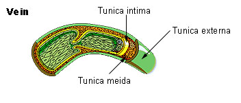 Vein
Vein -
 Anatomy of the arterial wall


External links
- Template:OklahomaHistology - "Aorta"
- Template:UCDavisOrganology - "Bird, vessels (LM, High)"
- Template:EMedicineDictionary
- Image at About.com