Acanthamoeba
| Acanthamoeba | ||||||
|---|---|---|---|---|---|---|
| Scientific classification | ||||||
|
Editor-In-Chief: C. Michael Gibson, M.S., M.D. [1]; Associate Editor(s)-in-Chief: Tamar Sifri [2]
Overview
Acanthamoeba is a genus of amoebae, one of the most common protozoa in soil, and also frequently found in fresh water and other habitats. The cells are small, usually 15 to 35 μm in length and oval to triangular in shape when moving. The pseudopods form a clear hemispherical lobe at the anterior, and there are various short filose extensions from the margins of the body. These give it a spiny appearance, which is what the name Acanthamoeba refers to. Cysts are common. Most species are free-living bacterivores, but some are opportunists that can cause infections in humans and other animals.
Acanthamoeba as a human pathogen
Diseases caused by Acanthamoeba include amoebic keratitis and encephalitis[1]. The latter is caused by Acanthamoeba entering cuts and spreading to the central nervous system. The former is a rare disease where amoebae invade the cornea of the eye. In the United States, it is nearly always associated with contact lens use, as Acanthamoeba can survive in the space between the lens and the eye.[2][3][4][5] However, elsewhere in the world, many cases of Acanthamoeba present in non-contact lens wearers.[6] For this reason, contact lenses must be properly disinfected before wearing, and should be removed when swimming or surfing.
To detect Acanthamoeba on a contact lens in a laboratory, a sheep blood agar plate with a layer (a lawn) of E. coli is made. Part of the contact lens is placed on the agar plate. If Acanthamoeba are present, they will ingest the bacteria, leaving a clear patch on the plate around the area of the lens. Polymerase chain reaction can also be used to confirm a diagnosis of Acanthamoeba keratitis, especially when contact lenses are not involved.[7]
Acanthamoeba and MRSA
Methicillin-resistant Staphylococcus aureus (MRSA) is an important pathogen in the hospital setting due to its resistance to many antibiotics. Recent findings demonstrate that MRSA can infect and replicate inside of Acanthamoeba polyphaga; this Acanthamoeba species is widespread throughout the environment. Since A. polyphaga can form cysts, cysts infected with MRSA can act as a mode of airborne dispersal for MRSA. Additionally, it is noted that "evidence with other pathogens suggests that pathogens that emerge from amoeba are more resistant to antibiotics and more virulent."[8] It has been observed that Acanthamoeba can increase MRSA numbers by 1000-fold.[9]
Importance of Acanthamoeba in soil ecology
A. castellanii can be found at high densities in various soil ecosystems. It preys on bacteria, but also fungi and other protozoa.
This species is able to lyse bacteria and produce a wide range of enzymes such as cellulases or chitinases[10] and probably contributes to the break down of organic matter in soil, contributing to the microbial loop.
Acanthamoeba species
Species of Acanthamoeba are distinguished mainly on the form of cysts, and include the following; those marked with an asterisk are known to cause infections.
A. astronyxis*
A. castellanii*
A. comandoni
A. culbertsoni*
A. divionensis
A. griffini
A. hatchetti*
A. healyi
A. jacobsi
A. lenticulata
A. lugdunensis*
A. mauritaniensis
A. palestinensis*
A. pearcei
A. polyphaga*
A. pustulosa
A. quina*
A. rhysodes*
A. royreba
A. terricola
A. triangularis
A. tubiashi
Endosymbiontes of Acanthamoeba
Acanthamoeba sp. contain diverse bacterial endosymbionts which are similar to human pathogens. Because of this they are considered to be potential emerging human pathogens.[11] The exact nature of these symbionts and the benefit they represent for the amoebal host still have to be clarified.
Life Cycle
Acanthamoeba spp. have been found in soil; fresh, brackish, and sea water; sewage; swimming pools; contact lens equipment; medicinal pools; dental treatment units; dialysis machines; heating, ventilating, and air conditioning systems; mammalian cell cultures; vegetables; human nostrils and throats; and human and animal brain, skin, and lung tissues. Unlike N. fowleri, Acanthamoeba has only two stages, cysts and trophozoites, in its life cycle. No flagellated stage exists as part of the life cycle. The trophozoites replicate by mitosis (nuclear membrane does not remain intact). The trophozoites are the infective forms, although both cysts and trophozoites gain entry into the body through various means. Entry can occur through the eye, the nasal passages to the lower respiratory tract, or ulcerated or broken skin. When Acanthamoeba spp. enters the eye it can cause severe keratitis in otherwise healthy individuals, particularly contact lens users. When it enters the respiratory system or through the skin, it can invade the central nervous system by hematogenous dissemination causing granulomatous amebic encephalitis (GAE) or disseminated disease, or skin lesions in individuals with compromised immune systems. Acanthamoeba spp. cysts and trophozoites are found in tissue.
-
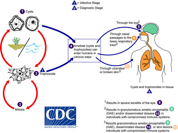 Acanthamoeba Life Cycle
Adapted from CDC

Medical Therapy
Antimicrobial Regimen
-
- Preferred regimen (1): Pentamidine AND Itraconazole AND Sulfadiazine AND Flucytosine
- Preferred regimen (2): Sulfadiazine AND Fluconazole AND Pyrimethamine
- Preferred regimen (3): Sulfadiazine AND Flucytosine AND TMP-SMX
- Preferred regimen (4): TMP-SMX AND Rifampin AND Ketoconazole
- Preferred regimen (5): Miltefosine AND Amikacin
- Preferred regimen (6): Miltefosine AND Voriconazole
- Preferred regimen (7): Pentamidine AND Itraconazole AND Flucytosine AND Levofloxacin AND Amphotericin B AND Rifampin
- Preferred regimen (8): Pentamidine AND Fluconazole AND Miltefosine
- Note: The mainstay of successful treatment includes early diagnosis and combination therapy with pentamidine, azole, sulfonamide, miltefosine, and possibly flucytosine.
- Preferred regimen: Pentamidine AND Sulfadiazine AND Flucytosine AND (Itraconazole OR Fluconazole) AND Chlorhexidine topical AND Ketoconazole topical
- 3. Acanthamoeba keratitis[17]
- Preferred regimen: (Polyhexamethylene biguanide topical OR Chlorhexidine topical) ± (Propamidine topical OR Hexamidine topical)
- Note (1): Azole antifungal drugs (Ketoconazole, Itraconazole, Voriconazole) may be considered as oral or topical adjuncts.
- Note (2): The duration of therapy for Acanthamoeba keratitis may last six months to a year.
- Note (3): Pain control can be helped by topical cyclopegic solutions and oral nonsteroidal medications.
- Note (4): The use of corticosteroids to control inflammation is controversial.
- Note (5): Penetrating keratoplasty may help restore visual acuity.
See also
- Legionella
- Momus (artist)
References
- Khan, N. A. (2006) Acanthamoeba: biology and increasing importance in human health. Fems Microbiology Reviews 30, 564-595.
- ↑ Di Gregorio, C (1992). "Acanthamoeba meningoencephalitis in a patient with acquired immunodeficiency syndrome". Archives of Pathology & Laboratory Medicine. 116 (12): 1363–5. PMID 1456885. Unknown parameter
|month=ignored (help); Unknown parameter|coauthors=ignored (help);|access-date=requires|url=(help) - ↑ Auran, JD (1987). "Acanthamoeba keratitis. A review of the literature". Cornea. 6 (1): 2–26. PMID 3556011. Unknown parameter
|coauthors=ignored (help);|access-date=requires|url=(help) - ↑ JOHN D.T. (1993) Opportunistically pathogenic free-living amebae. In: J.P. Kreier and J.R. Baker (Eds.), Parasitic Protozoa. Vol. 3. Academic Press, New York, pp. 143–246.
- ↑ Badenoch, PR (1995). "Corneal virulence, cytopathic effect on human keratocytes and genetic characterization of Acanthamoeba". International journal for parasitology. 25 (2): 229–39. PMID 7622330. Unknown parameter
|month=ignored (help); Unknown parameter|coauthors=ignored (help);|access-date=requires|url=(help) - ↑ Niederkorn, JY (1999). "The pathogenesis of Acanthamoeba keratitis". Microbes and Infection. 1 (6): 437–43. PMID 10602676. Unknown parameter
|month=ignored (help); Unknown parameter|coauthors=ignored (help);|access-date=requires|url=(help) - ↑ Sharma S, Garg P, Rao GN. "Patient characteristics, diagnosis, and treatment of non-contact lens related Acanthamoeba keratitis." The British Journal of Ophthalmology. 2000 Oct;84(10):1103-8. PMID: 11004092
- ↑ Pasricha, Gunisha (2003). "Use of 18S rRNA Gene-Based PCR Assay for Diagnosis of Acanthamoeba Keratitis in Non-Contact Lens Wearers in India". Journal of Clinical Microbiology. 41 (7): 3206–3211. doi: 10.1128/JCM.41.7.3206-3211.2003. Unknown parameter
|coauthors=ignored (help); Unknown parameter|month=ignored (help) - ↑ "MRSA use amoeba to spread, sidestepping hospital protection measures, new research shows" (Press release). University of Bath. 2006-02-28. Retrieved 2007-02-12.
- ↑ "Single Cell Amoeba Increases MRSA Numbers One Thousand Fold" (Press release). Blackwell Publishing. 2006-03-01. Retrieved 2007-02-12.
- ↑ Anderson, I. J. (2005). "Gene Discovery in the Acanthamoeba castellanii Genome". Protist. 156 (2): 203–14. PMID 16171187. Unknown parameter
|month=ignored (help); Unknown parameter|coauthors=ignored (help);|access-date=requires|url=(help) - ↑ Horn, M (2004). "Bacterial Endosymbionts of Free-living Amoebae". Journal of Eukaryotic Microbiology. 51 (5): 509–14. PMID 15537084. Unknown parameter
|month=ignored (help); Unknown parameter|coauthors=ignored (help);|access-date=requires|url=(help) - ↑ Visvesvara, Govinda S.; Moura, Hercules; Schuster, Frederick L. (2007-06). "Pathogenic and opportunistic free-living amoebae: Acanthamoeba spp., Balamuthia mandrillaris, Naegleria fowleri, and Sappinia diploidea". FEMS immunology and medical microbiology. 50 (1): 1–26. doi:10.1111/j.1574-695X.2007.00232.x. ISSN 0928-8244. PMID 17428307. Check date values in:
|date=(help) - ↑ Bennett, John (2015). Mandell, Douglas, and Bennett's principles and practice of infectious diseases. Philadelphia, PA: Elsevier/Saunders. ISBN 978-1455748013.
- ↑ Marciano-Cabral, Francine; Cabral, Guy (2003-04). "Acanthamoeba spp. as agents of disease in humans". Clinical Microbiology Reviews. 16 (2): 273–307. ISSN 0893-8512. PMC 153146. PMID 12692099. Check date values in:
|date=(help) - ↑ Bartlett, John (2012). Johns Hopkins ABX guide : diagnosis and treatment of infectious diseases. Burlington, MA: Jones and Bartlett Learning. ISBN 978-1449625580.
- ↑ Visvesvara, Govinda S.; Moura, Hercules; Schuster, Frederick L. (2007-06). "Pathogenic and opportunistic free-living amoebae: Acanthamoeba spp., Balamuthia mandrillaris, Naegleria fowleri, and Sappinia diploidea". FEMS immunology and medical microbiology. 50 (1): 1–26. doi:10.1111/j.1574-695X.2007.00232.x. ISSN 0928-8244. PMID 17428307. Check date values in:
|date=(help) - ↑ "Acanthamoeba Keratitis Fact Sheet (CDC)".
External links
- Acanthamoeba spp. as Agents of Disease in Humans - Clinical Microbiology Reviews, accessed on 4th February 2006
- Comprehensive resource on Amoeba -
- Eye health and Acanthamoeba