Coronary angiography right coronary artery: Difference between revisions
No edit summary |
|||
| (25 intermediate revisions by 3 users not shown) | |||
| Line 1: | Line 1: | ||
__NOTOC__ | __NOTOC__ | ||
{{Coronary angiography2}} | |||
{{CMG}} | {{CMG}} | ||
==Overview== | ==Overview== | ||
The right coronary artery (RCA) is a coronary artery with a single origin | The right coronary artery (RCA) is a coronary artery with a single origin near the right semilunar cusp of the [[aortic valve]] that bifurcates (branches) to supply the [[right ventricle|right ventricular]] free wall through the [[acute marginal branches]]), the inferior wall of the [[left ventricle]] through the [[posterior descending artery]], and the posterolateral wall of the [[left ventricle]] via the [[right posterolateral branch]]. On coronary angiography, the RCA is easily recognizable as it appears like the letter '''C''' in the left anterior oblique (LAO) projection and appears like a letter '''L''' in the right anterior oblique (RAO) projection. There are three angiographic views of the RCA that are traditionally obtained to visualize the proximal, middle, and distal segments. | ||
==How to Engage the Right Coronary Artery== | ==How to Engage the Right Coronary Artery== | ||
The | The right coronary artery (RCA) is engaged in the 30° LAO position. Using the [[femoral artery|femoral arterial]] approach, a Judkins Right 4 (JR4) catheter is traditionally used to engage the right coronary artery. The JR4 catheter is advanced into the body to make contact with the aortic valve. Next, the operator gently pulls the catheter out of the body about 2 cm while torquing the catheter clockwise. When the catheter faces to the left on the screen, it should be in or near the ostium of the right coronary artery. Other catheters that can be used to engage the right coronary artery include the Amplatz Right (AR1) and Amplatz Left (AL2 and AL3) catheters. If the origin of the right coronary artery has an upward trajectory, an internal mammary artery catheter may engage better. An inferiorly directed ostium may be cannulated with either a right coronary bypass catheter or an Amplatz catheter. | ||
==Optimal Views of the Right Coronary Artery== | ==Optimal Views of the Right Coronary Artery== | ||
The following sequence of views is obtained as the gantry is swung from the | The following sequence of 3 traditional views is obtained as the gantry is swung from the 30° LAO straight position to the AP 15° cranial position to the 30° RAO striaght position. | ||
===Proximal RCA=== | ===Proximal RCA=== | ||
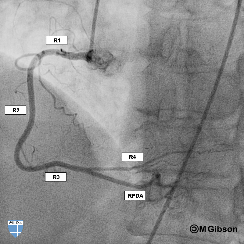
The | The Proximal RCA (R1 segment) including the ostium is best visualized in the 30° LAO view with no cranial or caudal angulation. | ||
{| | |||
|- | |||
| [[File:RCA_30_LAO.png|none|thumb|300px|]] | |||
| [[File:RCA_30_LAO.gif|none|thumb|300px|]] | |||
|- | |||
|} | |||
<span style="font-size:85%">R1 = Proximal right coronary artery; R2 = Middle right coronary artery; R3 = Distal right coronary artery; RPDA = Right posterior descending artery.</span> | |||
===Bifurcation of the RCA=== | ===Bifurcation of the RCA=== | ||
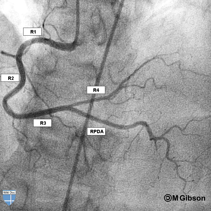
The bifurcation of the distal RCA where the [[right posterolateral artery]] and the [[posterior descending artery]] | The camera is next swung cranially to 15° - 20° and the LAO angulation is minimized to 5° - 10°. This view optimizes the bifurcation of the distal RCA where the [[right posterolateral artery]] and the [[posterior descending artery]] divide and branch from the distal right coronary artery. The patient should take a deep breath and hold it during the injection to optimize the view. | ||
{| | |||
|- | |||
| [[File:RCA_5_LAO_15_CRA.png|none|thumb|300px|]] | |||
| [[File:RCA_5_LAO_15_CRA.gif|none|thumb|300px|]] | |||
|- | |||
|} | |||
<span style="font-size:85%">R1 = Proximal right coronary artery; R2 = Middle right coronary artery; R3 = Distal right coronary artery; RPDA = Right posterior descending artery.</span> | |||
===Mid RCA=== | |||
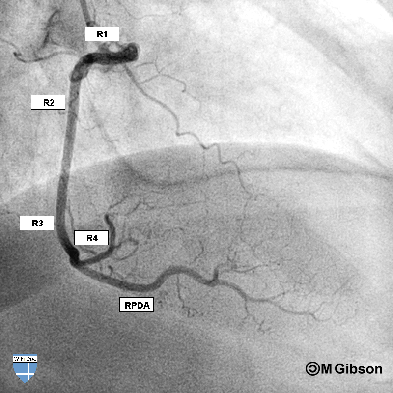
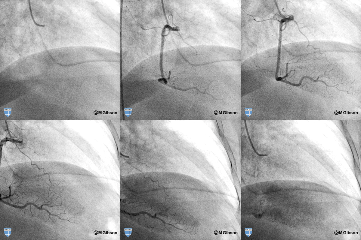
The middle RCA (R2 segment) is best visualized in the 30° RAO straight view. | |||
{| | {| | ||
|- | |- | ||
| [[ | | [[File:RCA_30_RAO.png|none|thumb|300px|]] | ||
| [[File:RCA_30_RAO_.gif|none|thumb|300px|]] | |||
|- | |- | ||
|} | |} | ||
=== | <span style="font-size:85%">R1 = Proximal right coronary artery; R2 = Middle right coronary artery; R3 = Distal right coronary artery; RPDA = Right posterior descending artery.</span> | ||
=== | ==Additional Images== | ||
==References== | ==References== | ||
{{Reflist|2}} | {{Reflist|2}} | ||
{{Arteries of chest}} | |||
{{Coronary Angiography}} | {{Coronary Angiography}} | ||
[[Category:Cardiology]] | |||
[[Category:Anatomy]] | |||
[[Category:Angiopedia]] | [[Category:Angiopedia]] | ||
[[Category:Arteries]] | |||
[[Category:Cardiac anatomy]] | |||
[[Category:Cardiology]] | [[Category:Cardiology]] | ||
[[Category:Cardiovascular system]] | |||
[[Category:Up-To-Date]] | |||
[[Category:Up-To-Date Cardiology]] | |||
{{WikiDoc Help Menu}} | |||
{{WikiDoc Sources}} | |||
Latest revision as of 15:17, 13 November 2013
|
Coronary Angiography | |
|
General Principles | |
|---|---|
|
Anatomy & Projection Angles | |
|
Normal Anatomy | |
|
Anatomic Variants | |
|
Projection Angles | |
|
Epicardial Flow & Myocardial Perfusion | |
|
Epicardial Flow | |
|
Myocardial Perfusion | |
|
Lesion Complexity | |
|
ACC/AHA Lesion-Specific Classification of the Primary Target Stenosis | |
|
Lesion Morphology | |
Editor-In-Chief: C. Michael Gibson, M.S., M.D. [1]
Overview
The right coronary artery (RCA) is a coronary artery with a single origin near the right semilunar cusp of the aortic valve that bifurcates (branches) to supply the right ventricular free wall through the acute marginal branches), the inferior wall of the left ventricle through the posterior descending artery, and the posterolateral wall of the left ventricle via the right posterolateral branch. On coronary angiography, the RCA is easily recognizable as it appears like the letter C in the left anterior oblique (LAO) projection and appears like a letter L in the right anterior oblique (RAO) projection. There are three angiographic views of the RCA that are traditionally obtained to visualize the proximal, middle, and distal segments.
How to Engage the Right Coronary Artery
The right coronary artery (RCA) is engaged in the 30° LAO position. Using the femoral arterial approach, a Judkins Right 4 (JR4) catheter is traditionally used to engage the right coronary artery. The JR4 catheter is advanced into the body to make contact with the aortic valve. Next, the operator gently pulls the catheter out of the body about 2 cm while torquing the catheter clockwise. When the catheter faces to the left on the screen, it should be in or near the ostium of the right coronary artery. Other catheters that can be used to engage the right coronary artery include the Amplatz Right (AR1) and Amplatz Left (AL2 and AL3) catheters. If the origin of the right coronary artery has an upward trajectory, an internal mammary artery catheter may engage better. An inferiorly directed ostium may be cannulated with either a right coronary bypass catheter or an Amplatz catheter.
Optimal Views of the Right Coronary Artery
The following sequence of 3 traditional views is obtained as the gantry is swung from the 30° LAO straight position to the AP 15° cranial position to the 30° RAO striaght position.
Proximal RCA
The Proximal RCA (R1 segment) including the ostium is best visualized in the 30° LAO view with no cranial or caudal angulation.
 |
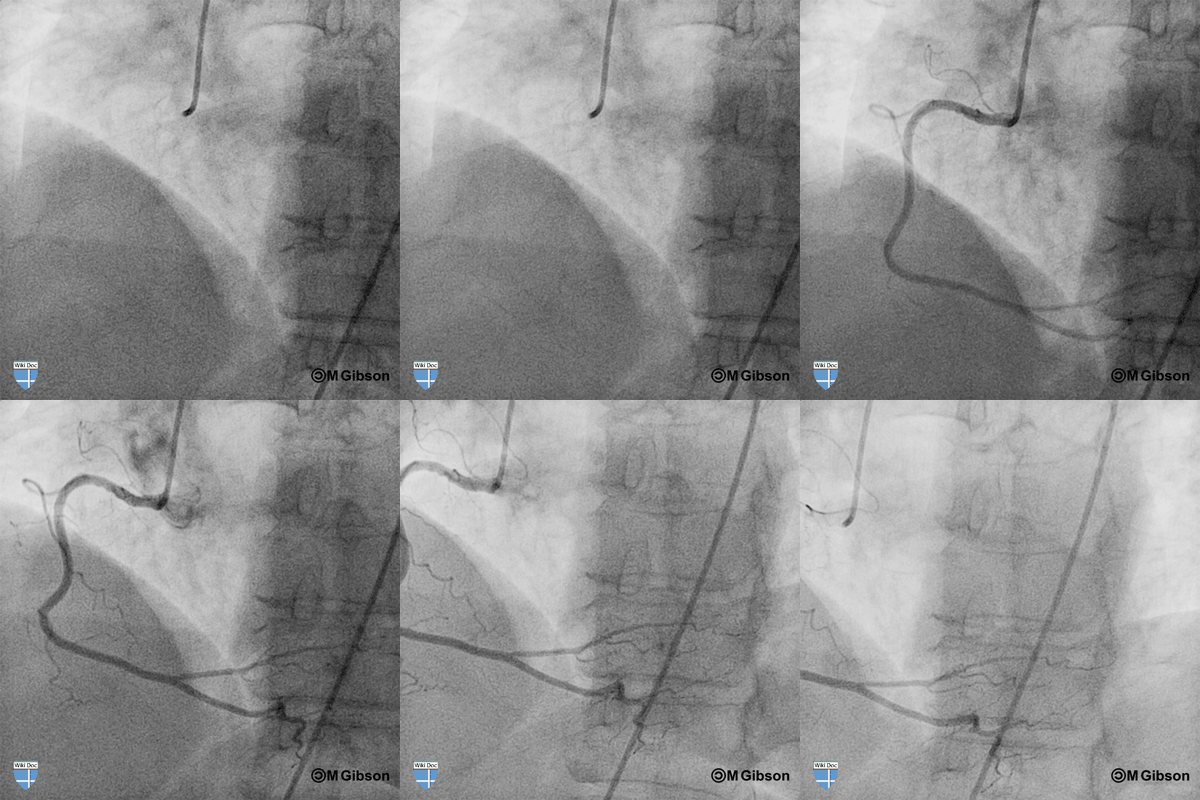 |
R1 = Proximal right coronary artery; R2 = Middle right coronary artery; R3 = Distal right coronary artery; RPDA = Right posterior descending artery.
Bifurcation of the RCA
The camera is next swung cranially to 15° - 20° and the LAO angulation is minimized to 5° - 10°. This view optimizes the bifurcation of the distal RCA where the right posterolateral artery and the posterior descending artery divide and branch from the distal right coronary artery. The patient should take a deep breath and hold it during the injection to optimize the view.
 |
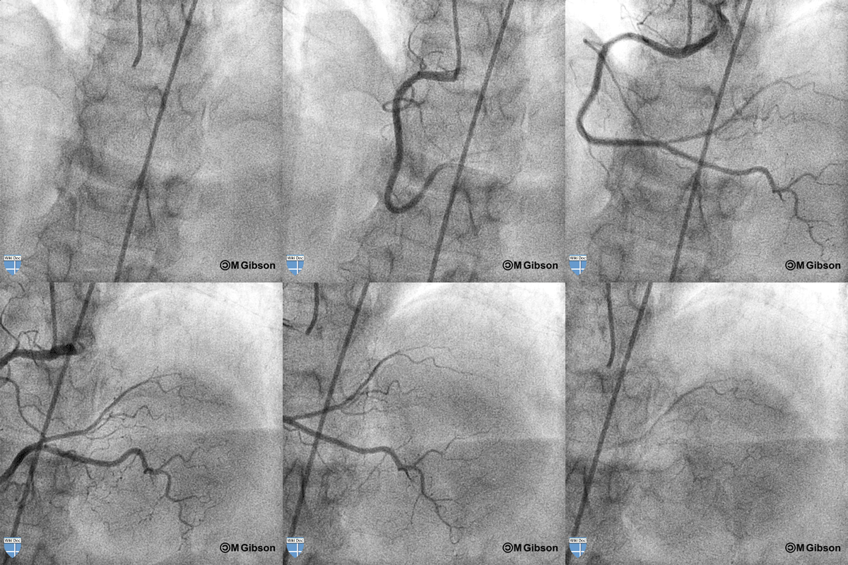 |
R1 = Proximal right coronary artery; R2 = Middle right coronary artery; R3 = Distal right coronary artery; RPDA = Right posterior descending artery.
Mid RCA
The middle RCA (R2 segment) is best visualized in the 30° RAO straight view.
 |
 |
R1 = Proximal right coronary artery; R2 = Middle right coronary artery; R3 = Distal right coronary artery; RPDA = Right posterior descending artery.