Femoral artery
Editor-In-Chief: C. Michael Gibson, M.S., M.D. [1]
Overview
The femoral artery is a large artery in the muscles of the thigh.
Structure
The femoral artery is a continuation of the external iliac artery, which comes from the abdominal aorta.
The external iliac artery becomes known as the femoral artery after it passes under the inguinal ligament. For a while at this location, (the femoral triangle), it is sometimes referred to as the common femoral, because it has not yet branched.
It usually gives off a branch known as the profunda femoris artery or the deep artery of the thigh, while continuing down the thigh medial to the femur. (The profunda femoris is even closer to the femur, and is more posterior).
The femoral artery goes through the adductor hiatus (a hole in the tendon of adductor magnus), into the posterior of the knee. Passing between the condyles of the femur, it becomes the popliteal artery of the popliteal fossa.
Branches
The femoral artery usually gives off the following branches:
- superficial epigastric artery
- Superficial circumflex iliac artery
- Superficial external pudendal artery
- Deep external pudendal artery
- Deep femoral artery
- Descending genicular artery
Clinical significance
The femoral artery pulse can be palpated at the femoral triangle.
Use of the term superficial femoral artery
Some specialist physicians (e.g. radiologists, vascular surgeons) call the femoral artery the superficial femoral artery after the profunda femoris artery branch point (to differentiate the femoral artery segments before and after the branch point). This term, historically, has not been used by anatomists and has fallen out of favour with most physicians because it has led to considerable confusion with its accompanying vein, the femoral vein, which if called superficial femoral vein might incorrectly be assumed to be a superficial vein, as opposed to a deep vein. (See article on femoral vein for more detailed discussion.)
Additional images
-
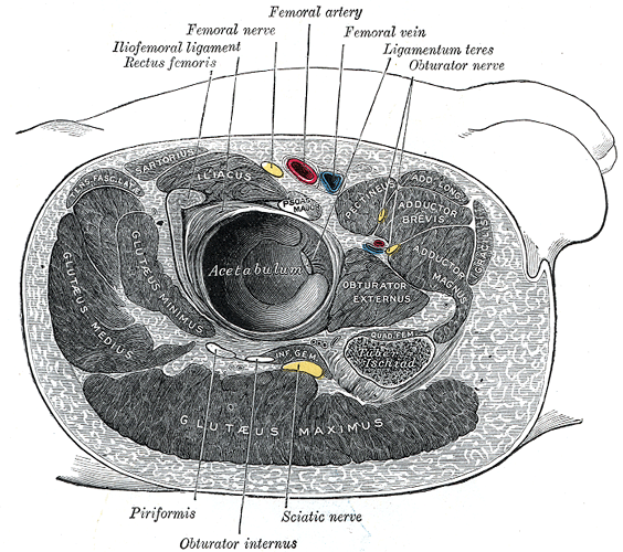 Structures surrounding right hip-joint.
 -
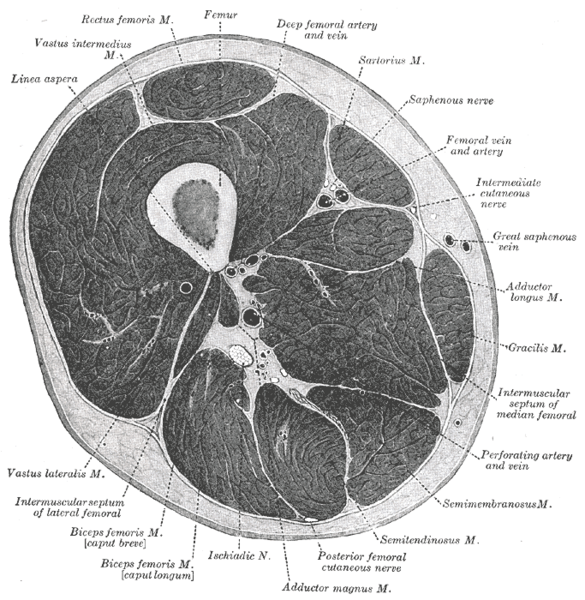 Cross-section through the middle of the thigh.
 -
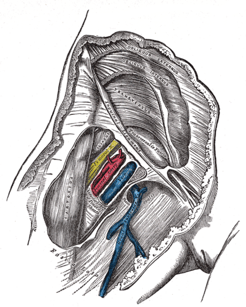 Femoral sheath laid open to show its three compartments.
 -
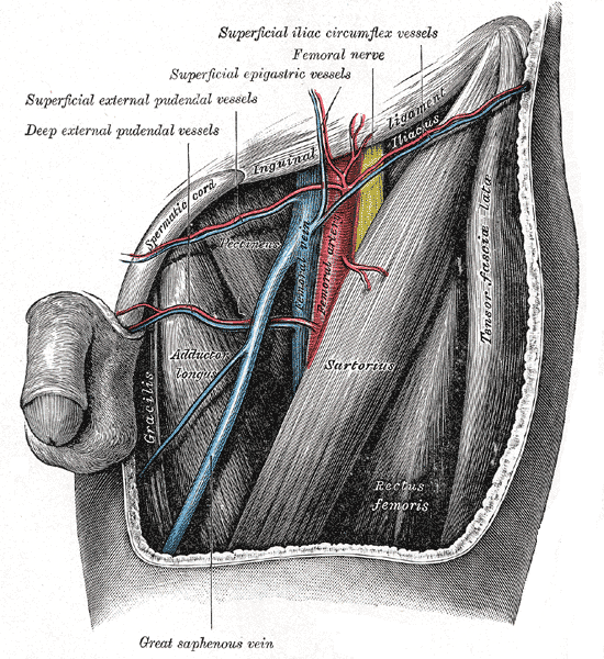 The left femoral triangle.
 -
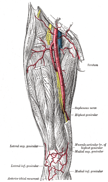 The femoral artery.
 -
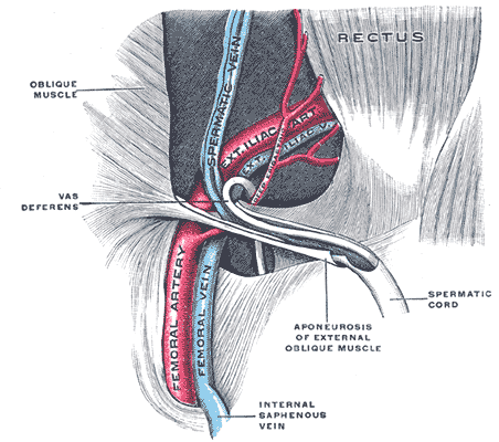 The spermatic cord in the inguinal canal.
 -
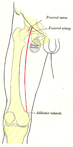 Front of right thigh, showing surface markings for bones, femoral artery and femoral nerve.


