* [[Vascular]] [[ischemia]] can lead to [[bowel]] [[congestion]] and result in decreased [[Absorption (digestive)|absorption]] of [[nutrients]] and can also cause [[ascites]] and more severe cases of [[mesenteric]] [[ischemia.]]<ref name="pmid7839976">{{cite journal |vauthors=Pantongrag-Brown L, Buetow PC, Carr NJ, Lichtenstein JE, Buck JL |title=Calcification and fibrosis in mesenteric carcinoid tumor: CT findings and pathologic correlation |journal=AJR Am J Roentgenol |volume=164 |issue=2 |pages=387–91 |date=February 1995 |pmid=7839976 |doi=10.2214/ajr.164.2.7839976 |url=}}</ref><ref name="pmid27861745">{{cite journal |vauthors=Daskalakis K, Karakatsanis A, Stålberg P, Norlén O, Hellman P |title=Clinical signs of fibrosis in small intestinal neuroendocrine tumours |journal=Br J Surg |volume=104 |issue=1 |pages=69–75 |date=January 2017 |pmid=27861745 |doi=10.1002/bjs.10333 |url=}}</ref>
* [[Vascular]] [[ischemia]] can lead to [[bowel]] [[congestion]] and result in decreased [[Absorption (digestive)|absorption]] of [[nutrients]] and can also cause [[ascites]] and more severe cases of [[mesenteric]] [[ischemia.]]<ref name="pmid7839976">{{cite journal |vauthors=Pantongrag-Brown L, Buetow PC, Carr NJ, Lichtenstein JE, Buck JL |title=Calcification and fibrosis in mesenteric carcinoid tumor: CT findings and pathologic correlation |journal=AJR Am J Roentgenol |volume=164 |issue=2 |pages=387–91 |date=February 1995 |pmid=7839976 |doi=10.2214/ajr.164.2.7839976 |url=}}</ref><ref name="pmid27861745">{{cite journal |vauthors=Daskalakis K, Karakatsanis A, Stålberg P, Norlén O, Hellman P |title=Clinical signs of fibrosis in small intestinal neuroendocrine tumours |journal=Br J Surg |volume=104 |issue=1 |pages=69–75 |date=January 2017 |pmid=27861745 |doi=10.1002/bjs.10333 |url=}}</ref>
==Genetics==
==Genetics==
*Gastrointestinal carcinoids occur in association with inherited syndromes, such as [[multiple endocrine neoplasia type 1]] and [[neurofibromatosis type 1]].<ref name="aaa">General Information About Gastrointestinal (GI) Carcinoid Tumors.<nowiki><ref name="pmid2886072"></nowiki>{{cite journal |vauthors=Duh QY, Hybarger CP, Geist R, Gamsu G, Goodman PC, Gooding GA, Clark OH |title=Carcinoids associated with multiple endocrine neoplasia syndromes |journal=Am. J. Surg. |volume=154 |issue=1 |pages=142–8 |date=July 1987 |pmid=2886072 |doi= |url=}}</ref>
*Gastrointestinal carcinoids occur in association with [[inherited syndromes]], such as [[multiple endocrine neoplasia type 1]] and [[neurofibromatosis type 1]].<ref name="aaa">General Information About Gastrointestinal (GI) Carcinoid Tumors.<nowiki><ref name="pmid2886072"></nowiki>{{cite journal |vauthors=Duh QY, Hybarger CP, Geist R, Gamsu G, Goodman PC, Gooding GA, Clark OH |title=Carcinoids associated with multiple endocrine neoplasia syndromes |journal=Am. J. Surg. |volume=154 |issue=1 |pages=142–8 |date=July 1987 |pmid=2886072 |doi= |url=}}</ref>
*[[Multiple endocrine neoplasia type 1]] is caused by alterations of the MEN1 gene located at chromosomal region 11q13.
*[[Multiple endocrine neoplasia type 1]] is caused by alterations of the [[MEN1]] [[gene]] located at [[chromosomal]] region [[11q13]].
*Most carcinoids associated with multiple endocrine neoplasia type 1 appear to be of foregut origin.
*Most [[Carcinoid|carcinoids]] associated with [[multiple endocrine neoplasia type 1]] appear to be of [[foregut]] origin.
*[[Neurofibromatosis type 1]] is an [[autosomal dominant]] genetic disorder caused by alteration of the ''[[NF1]]'' gene at chromosome 17q11.
*[[Neurofibromatosis type 1]] is an [[autosomal dominant]] [[genetic]] [[disorder]] caused by alteration of the ''[[NF1]]'' [[gene]] at [[chromosome]] [[17q11]].
*Carcinoids in patients with neurofibromatosis type 1 appear to arise primarily in the periampullary region.<ref name="pmid10873367">{{cite journal |vauthors=Karatzas G, Kouraklis G, Karayiannakis A, Patapis P, Givalos N, Kaperonis E |title=Ampullary carcinoid and jejunal stromal tumour associated with von Recklinghausen's disease presenting as gastrointestinal bleeding and jaundice |journal=Eur J Surg Oncol |volume=26 |issue=4 |pages=428–9 |date=June 2000 |pmid=10873367 |doi=10.1053/ejso.1999.0911 |url=}}</ref>
*[[Carcinoid|Carcinoids]] in patients with [[neurofibromatosis type 1]] appear to arise primarily in the [[periampullary]] region.<ref name="pmid10873367">{{cite journal |vauthors=Karatzas G, Kouraklis G, Karayiannakis A, Patapis P, Givalos N, Kaperonis E |title=Ampullary carcinoid and jejunal stromal tumour associated with von Recklinghausen's disease presenting as gastrointestinal bleeding and jaundice |journal=Eur J Surg Oncol |volume=26 |issue=4 |pages=428–9 |date=June 2000 |pmid=10873367 |doi=10.1053/ejso.1999.0911 |url=}}</ref>
* A hereditary form of small intestinal cacinoid tumour has been found which is caused by mutation in the IPMK gene leads to higher risk of developing carcinoid tumors in the small intestine.<ref name="pmid25865046">{{cite journal |vauthors=Sei Y, Zhao X, Forbes J, Szymczak S, Li Q, Trivedi A, Voellinger M, Joy G, Feng J, Whatley M, Jones MS, Harper UL, Marx SJ, Venkatesan AM, Chandrasekharappa SC, Raffeld M, Quezado MM, Louie A, Chen CC, Lim RM, Agarwala R, Schäffer AA, Hughes MS, Bailey-Wilson JE, Wank SA |title=A Hereditary Form of Small Intestinal Carcinoid Associated With a Germline Mutation in Inositol Polyphosphate Multikinase |journal=Gastroenterology |volume=149 |issue=1 |pages=67–78 |date=July 2015 |pmid=25865046 |pmc=4858647 |doi=10.1053/j.gastro.2015.04.008 |url=}}</ref>
* A [[hereditary]] form of [[small intestinal]] [[carcinoid]] [[Tumour|tumour has]] been found which is caused by [[Mutations|mutation]] in the IPMK [[gene]] leads to higher risk of developing [[carcinoid]] [[tumors]] in the [[Small intestinal|small intestine]].<ref name="pmid25865046">{{cite journal |vauthors=Sei Y, Zhao X, Forbes J, Szymczak S, Li Q, Trivedi A, Voellinger M, Joy G, Feng J, Whatley M, Jones MS, Harper UL, Marx SJ, Venkatesan AM, Chandrasekharappa SC, Raffeld M, Quezado MM, Louie A, Chen CC, Lim RM, Agarwala R, Schäffer AA, Hughes MS, Bailey-Wilson JE, Wank SA |title=A Hereditary Form of Small Intestinal Carcinoid Associated With a Germline Mutation in Inositol Polyphosphate Multikinase |journal=Gastroenterology |volume=149 |issue=1 |pages=67–78 |date=July 2015 |pmid=25865046 |pmc=4858647 |doi=10.1053/j.gastro.2015.04.008 |url=}}</ref>
*The most frequently reported mutated gene in gastrointestinal carcinoids is ''[[β-catenin]]'' (CTNNB1).<ref name="pmid11559529">{{cite journal |vauthors=Fujimori M, Ikeda S, Shimizu Y, Okajima M, Asahara T |title=Accumulation of beta-catenin protein and mutations in exon 3 of beta-catenin gene in gastrointestinal carcinoid tumor |journal=Cancer Res. |volume=61 |issue=18 |pages=6656–9 |date=September 2001 |pmid=11559529 |doi= |url=}}</ref>
*The most frequently reported [[mutated]] [[gene]] in [[gastrointestinal]] carcinoids is [[β-catenin|''β-catenin'']] (CTNNB1).<ref name="pmid11559529">{{cite journal |vauthors=Fujimori M, Ikeda S, Shimizu Y, Okajima M, Asahara T |title=Accumulation of beta-catenin protein and mutations in exon 3 of beta-catenin gene in gastrointestinal carcinoid tumor |journal=Cancer Res. |volume=61 |issue=18 |pages=6656–9 |date=September 2001 |pmid=11559529 |doi= |url=}}</ref>
==Embryology==
==Embryology==
*Carcinoid tumors originate from [[neuroendocrine cells]] ([[enterochromaffin]] or amine precursor uptake and decarboxylase [APUD] cells) which are derived from neural crest cells embrologically.
*[[Carcinoid tumors]] originate from [[neuroendocrine cells]] ([[enterochromaffin]] or [[amine]] precursor uptake and [[decarboxylase]] [[APUD cell|[APUD]]<nowiki>] </nowiki>[[cells]]) which are derived from [[neural crest cells]] [[embrologically]].
*Gastrointestinal carcinoids derive from cells that migrate from the neural crest to the [[foregut]], [[midgut]] and [[hindgut]].<ref name="pmid17114072">{{cite journal| author=Reznek RH| title=CT/MRI of neuroendocrine tumours. | journal=Cancer Imaging | year= 2006 | volume= 6 | issue= | pages= S163-77 | pmid=17114072 | doi=10.1102/1470-7330.2006.9037 | pmc=PMC1805060 | url=http://www.ncbi.nlm.nih.gov/entrez/eutils/elink.fcgi?dbfrom=pubmed&tool=sumsearch.org/cite&retmode=ref&cmd=prlinks&id=17114072 }} </ref>
*[[Gastrointestinal|Gastrointestina]]<nowiki/>l [[Carcinoid tumors|carcinoids]] derive from [[cells]] that migrate from the [[Neural crest cell|neural crest]] to the [[foregut]], [[midgut]] and [[hindgut]].<ref name="pmid17114072">{{cite journal| author=Reznek RH| title=CT/MRI of neuroendocrine tumours. | journal=Cancer Imaging | year= 2006 | volume= 6 | issue= | pages= S163-77 | pmid=17114072 | doi=10.1102/1470-7330.2006.9037 | pmc=PMC1805060 | url=http://www.ncbi.nlm.nih.gov/entrez/eutils/elink.fcgi?dbfrom=pubmed&tool=sumsearch.org/cite&retmode=ref&cmd=prlinks&id=17114072 }} </ref>
==Location==
==Location==
Carcinoid tumors are normally found throughout the gastrointestinal tract from [[mouth]] to [[anus]], with the highest concentration of cells in the [[appendix]] and [[small intestine]]. The [[pancreas]] contains a large number of these cells, the [[biliary tree]] only a few and the [[liver]] normally contains none. Fibrotic lesions are found on [[endocardium]], particularly on the right side of the [[heart]].
[[Carcinoid tumors]] are normally found throughout the [[gastrointestinal]] tract from [[mouth]] to [[anus]], with the highest concentration of [[cells]] in the [[appendix]] and [[small intestine]]. The [[pancreas]] contains a large number of these [[cells]], the [[biliary tree]] only a few and the [[liver]] normally contains none. [[Fibrosis|Fibrotic]] lesions are found on [[endocardium]], particularly on the right side of the [[heart]].
==Gross Pathology==
==Gross Pathology==
===Gastrointestinal Carcinoid===
===Gastrointestinal Carcinoid===
In the gastric or intestinal wall, carcinoids may occur as firm white, yellow, or gray nodules and may be intramural masses or may protrude into the lumen as polypoid nodules. The overlying [[gastric]] or [[intestinal mucosa]] may be intact or have focal ulceration.
In the [[gastric]] or [[intestinal]] wall, [[Carcinoid tumors|carcinoids tumoors]] may occur as firm white, yellow, or gray nodules and may be [[intramural masses]] or may protrude into the [[lumen]] as polypoid [[nodules]]. The overlying [[gastric]] or [[intestinal mucosa]] may be intact or have focal [[ulceration]].
*Well-differentiated NETs of the tubular gastrointestinal tract are often well-circumscribed round lesions in the submucosa or extending to the muscular layer.
*Well-differentiated [[neuroendocrine tumors]] of the [[tubular]] [[gastrointestinal tract]] are often [[Circumscribed|well-circumscribed]] round lesions in the [[Submucosal|submucosa]] or extending to the [[Muscular|muscular layer]].
*The cut surface appears red to tan, reflecting the abundant microvasculature, or sometimes yellow because of high lipid content.
*The cut surface appears red to tan, reflecting the abundant [[microvasculature]], or sometimes yellow because of high [[lipid]] content.
{| align=""
{| align=""
Line 77:
Line 77:
===Neuroendocrine tumours of the lung===
===Neuroendocrine tumours of the lung===
*Pulmonary neoplasms that are characterized by neuroendocrine differentiation and relatively indolent clinical behavior.
*[[Pulmonary]] [[neoplasms]] that are characterized by [[Neuroendocrine|neuroendocrin]]<nowiki/>e differentiation and relatively indolent [[clinical]] behavior.
*Lung is the second most common site for neuroendocrine tumours.
*Lung is the second most common site for [[neuroendocrine tumor]].
*Lung NET are classified on the basis of histology:
*[[Lung]] [[Neuroendocrine tumor|neuroendocrine tumors]] are classified on the basis of [[Histology|histology:]]
#Typical neuroendocrine tumours :well-differentiated, low-grade, slowly growing neoplasms that seldom metastasize to extrathoracic structures and localized.
#Typical [[neuroendocrine tumor]] :well-differentiated, low-grade, slowly growing [[neoplasms]] that seldom [[metastasize]] to [[extrathoracic]] structures and localized.
#Poorly differentiated and high-grade neuroendocrine carcinomas, as typified by small cell lung cancer and large cell carcinomas which behaves aggressively, with rapid tumor growth and early distant dissemination.
#Poorly [[differentiated]] and high-grade [[Neuroendocrine tumor|neuroendocrine carcinomas]], as typified by [[small cell lung cancer]] and [[large cell carcinomas]] which behaves [[aggressively]], with rapid [[tumor]] growth and early distant [[dissemination]].
#Atypical neuroendorcrine tumors, which are of intermediate grade and differentiation, is intermediate between typical neuroendocrine tumors and small cell lung cancer.
#Atypical [[neuroendocrine tumor]], which are of intermediate grade and differentiation, is intermediate between typical [[neuroendocrine tumor]] and [[small cell lung cancer]].
*Based on the location:
*Based on the location:
Carcinoid tumor of the lung may be classified based on the location into two subtypes:
[[Carcinoid tumor]] of the [[lung]] may be classified based on the location into two subtypes:
#Bronchial carcinoid tumors: central lesions
#[[Bronchial]] [[carcinoid tumors]]: central lesions
*NETs arise from enterochromaffin (neuroendocrine) cells of the aerodigstive tract.
*[[Neuroendocrine tumor]] arise from [[Enterochromaffin cells|enterochromaffin]] ([[Neuroendocrine cells|neuroendocrine) cells]] of the [[Gastrointestinal|gastrointestinal tract.]]
*The term enterochromaffin refers to the ability to stain with potassium chromate (chromaffin), a feature of cells that contain serotonin.
*The term [[enterochromaffin]] refers to the ability to stain with [[Potassium chromate|potassium chromate (chromaffin]]), a feature of [[cells]] that contain [[serotonin]].
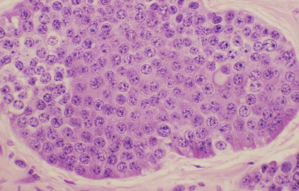
* On electron microscopy ,the cells in tumors are found to contain membrane-bound secretory granules with dense-core granules in the cytoplasm.[[File:Typical carcinoid histopathology.jpg|thumb|left|700px| Typical carcinoid histopathology-The nuclei of the tumor cells are uniform with a stippled chromatin pattern. There is no mitotic activity or necrosis. https://commons.wikimedia.org/wiki/File:Typical_carcinoid_(3931156341).jpg source Case courtesy of Dr Yale Rosen, from wikicommons]]
* On [[electron microscopy]] ,the [[cells]] in [[tumors]] are found to contain [[membrane]]-bound [[Granules|secretory granules]] with dense-core [[granules]] in the [[cytoplasm]].[[File:Typical carcinoid histopathology.jpg|thumb|left|700px| Typical carcinoid histopathology-The nuclei of the tumor cells are uniform with a stippled chromatin pattern. There is no mitotic activity or necrosis. https://commons.wikimedia.org/wiki/File:Typical_carcinoid_(3931156341).jpg source Case courtesy of Dr Yale Rosen, from wikicommons]]The most recent nomenclature for [[neuroendocrine tumor]]<nowiki/>s of the [[Digestive systems|digestive system]] from the [[World Health Organization]] (WHO) distinguishes two broad subgroups:<ref name="pmid21629514">{{cite journal |vauthors=Schott M, Klöppel G, Raffel A, Saleh A, Knoefel WT, Scherbaum WA |title=Neuroendocrine neoplasms of the gastrointestinal tract |journal=Dtsch Arztebl Int |volume=108 |issue=18 |pages=305–12 |date=May 2011 |pmid=21629514 |pmc=3103981 |doi=10.3238/arztebl.2011.0305 |url=}}</ref><ref name="pmid28837143">{{cite journal |vauthors=Cavalcanti E, Armentano R, Valentini AM, Chieppa M, Caruso ML |title=Role of PD-L1 expression as a biomarker for GEP neuroendocrine neoplasm grading |journal=Cell Death Dis |volume=8 |issue=8 |pages=e3004 |date=August 2017 |pmid=28837143 |pmc=5596583 |doi=10.1038/cddis.2017.401 |url=}}</ref><ref name="pmid25412850">{{cite journal |vauthors=Reid MD, Bagci P, Ohike N, Saka B, Erbarut Seven I, Dursun N, Balci S, Gucer H, Jang KT, Tajiri T, Basturk O, Kong SY, Goodman M, Akkas G, Adsay V |title=Calculation of the Ki67 index in pancreatic neuroendocrine tumors: a comparative analysis of four counting methodologies |journal=Mod. Pathol. |volume=28 |issue=5 |pages=686–94 |date=May 2015 |pmid=25412850 |pmc=4460192 |doi=10.1038/modpathol.2014.156 |url=}}</ref>
*The most recent nomenclature for NETs of the digestive system from the World Health Organization (WHO) distinguishes two broad subgroups:<ref name="pmid21629514">{{cite journal |vauthors=Schott M, Klöppel G, Raffel A, Saleh A, Knoefel WT, Scherbaum WA |title=Neuroendocrine neoplasms of the gastrointestinal tract |journal=Dtsch Arztebl Int |volume=108 |issue=18 |pages=305–12 |date=May 2011 |pmid=21629514 |pmc=3103981 |doi=10.3238/arztebl.2011.0305 |url=}}</ref><ref name="pmid28837143">{{cite journal |vauthors=Cavalcanti E, Armentano R, Valentini AM, Chieppa M, Caruso ML |title=Role of PD-L1 expression as a biomarker for GEP neuroendocrine neoplasm grading |journal=Cell Death Dis |volume=8 |issue=8 |pages=e3004 |date=August 2017 |pmid=28837143 |pmc=5596583 |doi=10.1038/cddis.2017.401 |url=}}</ref><ref name="pmid25412850">{{cite journal |vauthors=Reid MD, Bagci P, Ohike N, Saka B, Erbarut Seven I, Dursun N, Balci S, Gucer H, Jang KT, Tajiri T, Basturk O, Kong SY, Goodman M, Akkas G, Adsay V |title=Calculation of the Ki67 index in pancreatic neuroendocrine tumors: a comparative analysis of four counting methodologies |journal=Mod. Pathol. |volume=28 |issue=5 |pages=686–94 |date=May 2015 |pmid=25412850 |pmc=4460192 |doi=10.1038/modpathol.2014.156 |url=}}</ref>
*Well-differentiated [[Neuroendocrine tumor|neuroendocrine tumor:]] which are further subdivided according to proliferative rate:
*Well-differentiated NETs: which are further subdivided according to proliferative rate:
#Low grade
#Low grade
#Intermediate grade.(Intermediate-grade NETs arising in the lung (but not elsewhere) are referred to as atypical carcinoids
#Intermediate grade.(Intermediate-grade [[neuroendocrine tumor]] arising in the [[lung]] (but not elsewhere) are referred to as atypical [[Carcinoid|carcinoid.]]
*They are high-grade carcinomas that resemble small cell or large cell neuroendocrine carcinoma of the lung.
*They are high-grade [[carcinomas]] that resemble [[Small cell carcinoma of the lung|small cell carcinoma]] or [[large cell neuroendocrine carcinoma of the lung]].
*Histoloigically, well-differentiated NETs have characteristic "organoid" arrangements of tumor cells, with solid/nesting, trabecular, gyriform, or sometimes, glandular patterns.
*Histoloigically, well-differentiated [[neuroendocrine tumor]] have characteristic "[[organoid]]" arrangements of [[Tumor cell|tumor]] [[cells]], with solid/nesting, trabecular, [[gyriform]], or sometimes, [[glandular]] patterns.
*The cells are relatively uniform, and they have round to oval nuclei, coarsely stippled chromatin, and finely granular cytoplasm.
*The [[cells]] are relatively uniform, and they have round to oval nuclei, coarsely stippled [[Chromatin|chromatin,]] and finely [[Granularity|granular]] [[Cytoplasmic|cytoplasm]].
*The cells produce abundant neurosecretory granules, as reflected in the strong and diffuse immunohistochemical expression of neuroendocrine markers such as synaptophysin and chromogranin.
*The [[cells]] produce abundant [[Neurosecretory|neurosecretory granules]], as reflected in the strong and diffuse [[immunohistochemical]] expression of [[Neuroendocrine cells|neuroendocrine markers]] such as [[synaptophysin]] and [[chromogranin]].
*Well-differentiated NETs of the midgut (ileum in particular) also have a very characteristic pattern of solid or cribriform nests punctuated by sharply outlined luminal spaces with peripheral nuclear palisading and granular eosinophilic cytoplasm.
*Well-differentiated [[neuroendocrine tumor]] of the [[midgut]] [[Ileum|(ileum]] in particular) also have a very characteristic pattern of [[solid]] or cribriform nests punctuated by sharply outlined luminal spaces with peripheral [[nuclear]] [[palisading]] and [[Granular cell|granular]] [[eosinophilic]] [[Cytoplasmic|cytoplasm]].
*Poorly differentiated neuroendocrine carcinomas (NECs) less closely resemble nonneoplastic neuroendocrine cells and have a more sheet-like or diffuse architecture, irregular nuclei, and less cytoplasmic granularity. Immunohistochemical expression of neuroendocrine markers is generally more limited in extent and intensity.
*Poorly differentiated [[Neuroendocrine tumor|neuroendocrine carcinomas]] (NECs) less closely resemble [[nonneoplastic]] [[neuroendocrine cells]] and have a more sheet-like or diffuse architecture, irregular [[nuclei]], and less [[cytoplasmic]] [[granularity]]. [[Immunohistochemical|Immunohistochemica]]<nowiki/>l expression of [[Neuroendocrine cells|neuroendocrine]] markers is generally more limited in extent and intensity.
Left upper lobe": A lung lobe 185x110x55mm with bronchovascular remnants up to 25mm. Arising in the hilum and involving the bronchus is a rubbery tan-pink tumor 21x20x19mm. The tumor is 6mm from the bronchovascular margins and 3mm from the hilar margin. 26mm from the tumor and 1mm from the pleura there is a firm white nodule 6mm. Peripheral to the tumor is an area where the lung shows dilated bronchi up to 12mm in diameter which lie 2mm from the pleura.Source: Radiopedia
↑Kvols LK, Moertel CG, O'Connell MJ, Schutt AJ, Rubin J, Hahn RG (September 1986). "Treatment of the malignant carcinoid syndrome. Evaluation of a long-acting somatostatin analogue". N. Engl. J. Med. 315 (11): 663–6. doi:10.1056/NEJM198609113151102. PMID2427948.
↑Grozinsky-Glasberg S, Grossman AB, Gross DJ (2015). "Carcinoid Heart Disease: From Pathophysiology to Treatment--'Something in the Way It Moves'". Neuroendocrinology. 101 (4): 263–73. doi:10.1159/000381930. PMID25871411.
↑Launay JM, Birraux G, Bondoux D, Callebert J, Choi DS, Loric S, Maroteaux L (February 1996). "Ras involvement in signal transduction by the serotonin 5-HT2B receptor". J. Biol. Chem. 271 (6): 3141–7. PMID8621713.
↑Xu J, Jian B, Chu R, Lu Z, Li Q, Dunlop J, Rosenzweig-Lipson S, McGonigle P, Levy RJ, Liang B (December 2002). "Serotonin mechanisms in heart valve disease II: the 5-HT2 receptor and its signaling pathway in aortic valve interstitial cells". Am. J. Pathol. 161 (6): 2209–18. doi:10.1016/S0002-9440(10)64497-5. PMID12466135.
↑Luis SA, Pellikka PA (January 2016). "Carcinoid heart disease: Diagnosis and management". Best Pract. Res. Clin. Endocrinol. Metab. 30 (1): 149–58. doi:10.1016/j.beem.2015.09.005. PMID26971851.
↑Druce MR, Bharwani N, Akker SA, Drake WM, Rockall A, Grossman AB (March 2010). "Intra-abdominal fibrosis in a recent cohort of patients with neuroendocrine ('carcinoid') tumours of the small bowel". QJM. 103 (3): 177–85. doi:10.1093/qjmed/hcp191. PMID20123681.
↑Pantongrag-Brown L, Buetow PC, Carr NJ, Lichtenstein JE, Buck JL (February 1995). "Calcification and fibrosis in mesenteric carcinoid tumor: CT findings and pathologic correlation". AJR Am J Roentgenol. 164 (2): 387–91. doi:10.2214/ajr.164.2.7839976. PMID7839976.
↑Daskalakis K, Karakatsanis A, Stålberg P, Norlén O, Hellman P (January 2017). "Clinical signs of fibrosis in small intestinal neuroendocrine tumours". Br J Surg. 104 (1): 69–75. doi:10.1002/bjs.10333. PMID27861745.
↑General Information About Gastrointestinal (GI) Carcinoid Tumors.<ref name="pmid2886072">Duh QY, Hybarger CP, Geist R, Gamsu G, Goodman PC, Gooding GA, Clark OH (July 1987). "Carcinoids associated with multiple endocrine neoplasia syndromes". Am. J. Surg. 154 (1): 142–8. PMID2886072.
↑Karatzas G, Kouraklis G, Karayiannakis A, Patapis P, Givalos N, Kaperonis E (June 2000). "Ampullary carcinoid and jejunal stromal tumour associated with von Recklinghausen's disease presenting as gastrointestinal bleeding and jaundice". Eur J Surg Oncol. 26 (4): 428–9. doi:10.1053/ejso.1999.0911. PMID10873367.
↑Fujimori M, Ikeda S, Shimizu Y, Okajima M, Asahara T (September 2001). "Accumulation of beta-catenin protein and mutations in exon 3 of beta-catenin gene in gastrointestinal carcinoid tumor". Cancer Res. 61 (18): 6656–9. PMID11559529.
.JPG)


.jpg)