Arteriovenous malformation
| Arteriovenous malformation | |
 | |
|---|---|
| Brain: Arteriovenous Malformation: Gross fixed tissue close-up view of malformation in meninges and cerebral cortex (an excellent example). Image courtesy of Professor Peter Anderson DVM PhD and published with permission © PEIR, University of Alabama at Birmingham, Department of Pathology |
|
Arteriovenous malformations Microchapters |
|
Differentiating Arteriovenous malformations from other Diseases |
|---|
|
Diagnosis |
|
Treatment |
|
Case Studies |
|
Arteriovenous malformation On the Web |
|
American Roentgen Ray Society Images of Arteriovenous malformation |
|
Directions to Hospitals Treating Arteriovenous malformations |
|
Risk calculators and risk factors for Arteriovenous malformation |
Editor-In-Chief: C. Michael Gibson, M.S., M.D. [1]; Associate Editor(s)-in-Chief: Kalsang Dolma, M.B.B.S.[2]
Synonyms and keywords: AVM; arterio-venous malformations
Overview
Arteriovenous malformation is a congenital disorder of the connections between veins and arteries in the vascular system. The genetic transmission patterns of AVM (if any) are unknown, and AVM is not generally thought to be an inherited disorder--unless in the context of a specific hereditary syndrome.
Pathophysiology
Arteries and veins are part of the human cardiovascular system. Normally, the arteries in the vascular system carry oxygen-rich blood at a relatively high pressure. Structurally, arteries divide and sub-divide repeatedly, eventually forming a sponge-like capillary bed. Blood moves through the capillaries, giving up oxygen and taking up waste products from the surrounding cells. Capillaries successively join together, one upon the other, to form the veins that carry blood away at a relatively low pressure. The heart acts to pump blood from the low pressure veins to the high pressure arteries.
If the capillary bed is thought of as a sponge, then an AVM is the rough equivalent of inserting a tangle of drinking straw|flexible soda straws from artery to vein through that sponge. On an arteriogram, an AVM often resemble a tangle of spaghetti noodles. This tangle of blood vessels forms a relatively direct connection between high pressure arteries and low pressure veins and bypasses the capillary bed.
The result is a collection of blood vessels with abnormal connections and no capillaries. This collection, often called a nidus, can be extremely fragile and prone to bleeding. This bleeding can be catastrophic if it occurs in the brain, the lung or the gastrointestinal tract.
AVMs can occur in various parts of the body
- Brain, causing a cerebral arteriovenous malformation
- Spleen[1]
- Lung[2]
- Kidney[3]
- Spinal cord[4]
- Liver[5]
- Intercostal space[6]
- Iris[7]
- Spermatic cord[8]
Genetics
Can occur due to autosomal dominant diseases, such as Hereditary Hemorrhagic Telangiectasia
Epidemiology and Demographics
An estimated 300,000 Americans have AVMs, of which 12% (approximately 36,000) will exhibit symptoms that differ greatly in severity.
Diagnosis
Symptoms
Symptoms of AVM vary according to the location of the malformation. Roughly (88% -needs citation) AVM are asymptomatic; often the malformation is discovered as part of an autopsy or during treatment of an unrelated disorder (called in medicine an incidental finding), rarely its expansion or a micro-bleed from it, could cause epilepsy, deficit or elicit pain. The most general symptoms include
- Headache
- Epilepsy,
- Difficulties with movement or coordination, including muscle weakness and even paralysis;
- vertigo (dizziness);
- Difficulties of speech (dysarthria) and communication, such as alogia;
- Difficulties with everyday activities, such as apraxia;
- Abnormal sensations (numbness, tingling, or spontaneous pain);
- Memory and thought-related problems, such as confusion, dementia or hallucinations.
Patient #1: MRI demonstrates a large right AVM
-
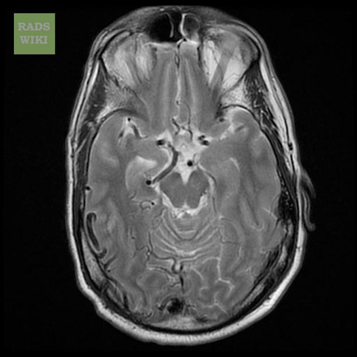
T2
-
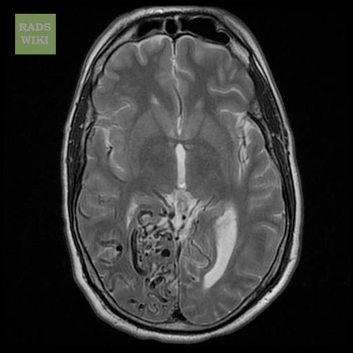
T2
-
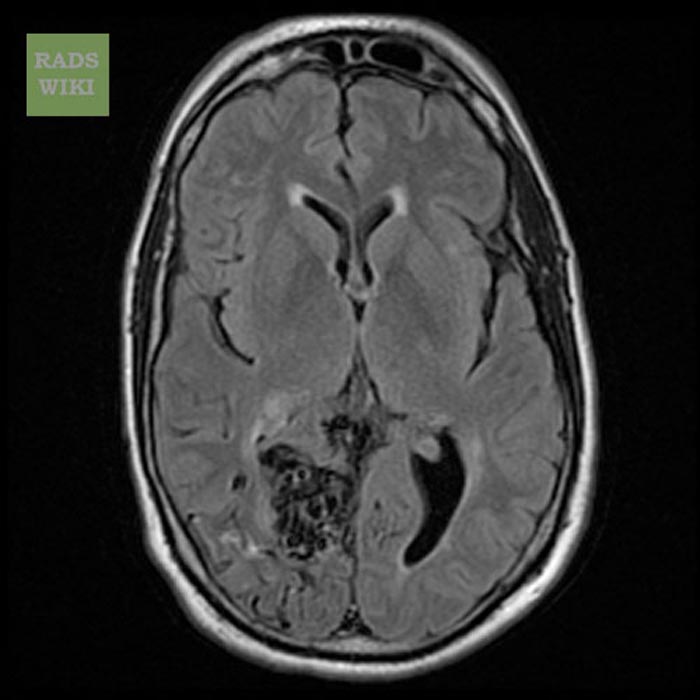
FLAIR
-
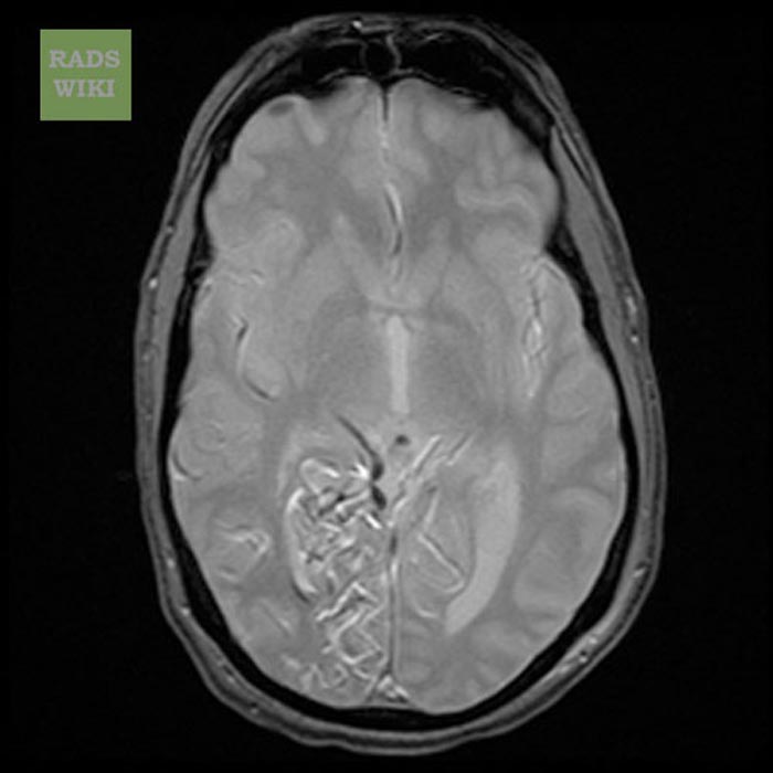
GRE
-
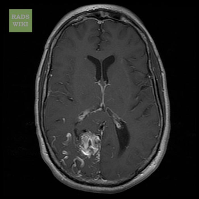
T1 with GAD
-
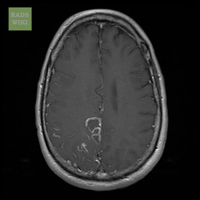
T1 with GAD
-
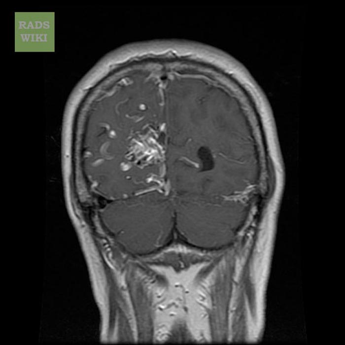
T1 with GAD
-
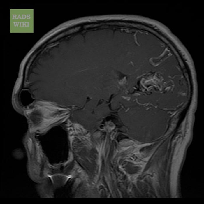
T1 with GAD


Treatment
Treatment can be symptomatic, or it can involve surgery or radiation therapy.
It can also be carried out via in interventinal radiography procedure using a glue to cut off the blood supply to the AVM.
Research directions
The optimal management of AVMs remains an ongiong topic of clinical research. [9].
References
- ↑ Agrawal A, Whitehouse R, Johnson RW, Augustine T (2006). "Giant splenic artery aneurysm associated with arteriovenous malformation". J. Vasc. Surg. 44 (6): 1345–9. doi:10.1016/j.jvs.2006.06.049. PMID 17145440. Retrieved 2008-06-01. Unknown parameter
|month=ignored (help) - ↑ Chowdhury UK, Kothari SS, Bishnoi AK, Gupta R, Mittal CM, Reddy S (2008). "Successful Lobectomy for Pulmonary Arteriovenous Malformation Causing Recurrent Massive Haemoptysis". Heart Lung Circ. doi:10.1016/j.hlc.2007.11.142. PMID 18294908. Retrieved 2008-06-01. Unknown parameter
|month=ignored (help) - ↑ Barley FL, Kessel D, Nicholson T, Robertson I (2006). "Selective embolization of large symptomatic iatrogenic renal transplant arteriovenous fistula". Cardiovasc Intervent Radiol. 29 (6): 1084–7. doi:10.1007/s00270-005-0265-z. PMID 16794894.
- ↑ Kishi K, Shirai S, Sonomura T, Sato M (2005). "Selective conformal radiotherapy for arteriovenous malformation involving the spinal cord". Br J Radiol. 78 (927): 252–4. PMID 15730991. Unknown parameter
|month=ignored (help) - ↑ Bauer T, Britton P, Lomas D, Wight DG, Friend PJ, Alexander GJ (1995). "Liver transplantation for hepatic arteriovenous malformation in hereditary haemorrhagic telangiectasia". J. Hepatol. 22 (5): 586–90. PMID 7650340. Retrieved 2008-06-01. Unknown parameter
|month=ignored (help) - ↑ Rivera PP, Kole MK, Pelz DM, Gulka IB, McKenzie FN, Lownie SP (2006). "Congenital intercostal arteriovenous malformation". AJR Am J Roentgenol. 187 (5): W503–6. doi:10.2214/AJR.05.0367. PMID 17056881. Unknown parameter
|month=ignored (help) - ↑ Shields JA, Streicher TF, Spirkova JH, Stubna M, Shields CL (2006). "Arteriovenous malformation of the iris in 14 cases". Arch. Ophthalmol. 124 (3): 370–5. doi:10.1001/archopht.124.3.370. PMID 16534057. Unknown parameter
|month=ignored (help) - ↑ Sountoulides P, Bantis A, Asouhidou I, Aggelonidou H (2007). "Arteriovenous malformation of the spermatic cord as the cause of acute scrotal pain: a case report". J Med Case Reports. 1: 110. doi:10.1186/1752-1947-1-110. PMID 17939869.
- ↑ Research trials in arterio-venous malformations; Rustam Al-Shahi Salman