Iris (anatomy)
Template:Infobox Anatomy Editor-In-Chief: C. Michael Gibson, M.S., M.D. [1]
Overview
In anatomy, the iris (plural irises or irides) is the most visible part of the eye of vertebrates, including humans. The following describes the iris of vertebrates, not the independently evolved iris found in some cephalopods. The word comes from Greek mythology, in which Iris is the anthropomorphized form of the rainbow.
The iris consists of pigmented fibrovascular tissue known as a stroma. The stroma connects a sphincter muscle (sphincter pupillae), which contracts the pupil, and a set of dilator muscles (dilator pupillae) which open it. The back surface is covered by an epithelial layer two cells thick (the iris pigment epithelium), but the front surface has no epithelium. The outer edge of the iris, known as the root, is attached to the sclera and the anterior ciliary body. The iris and ciliary body together are known as the anterior uvea. Just in front of the root of the iris is the region through which the aqueous humour constantly drains out of the eye, with the result that diseases of the iris often have important effects on intraocular pressure, and indirectly on vision.
General structure
The iris is divided into two major regions:
- The pupillary zone is the inner region whose edge forms the boundary of the pupil.
- The ciliary zone is the rest of the iris that extends to its origin at the ciliary body.
The collarette is the region of the iris separating the pupillary portion from the ciliary portion. It is typically defined as the region where the sphincter muscle and dilator muscle overlap.
Histological features
From anterior (front) to posterior (back), the layers of the iris are:
- Anterior border layer
- Stroma of iris
- Iris sphincter muscle
- Iris dilator muscle
- Anterior pigment myoepithelium
- Posterior pigment epithelium
Anterior surface features
- The Crypts of Fuchs are a series of openings located on either side of the collarette that allow the stroma and deeper iris tissues to be bathed in aqueous humor. Collagen trabeculae that surround the border of the crypts can be seen in blue irises.
- The pupillary ruff is a series of small ridges at the pupillary margin formed by the continuation of the pigmented epithelium from the posterior surface.
- The Circular contraction folds, also known as contraction furrows, are a series of circular bands or folds about midway between the collarette and the origin of the iris. These folds result from changes in the surface of the iris as it dilates.
- Crypts at the base of the iris are additional openings that can be observed close to the outermost part of the ciliary portion of the iris.
Posterior surface features
- The Radial contraction folds of Schwalbe are a series of very fine radial folds in the pupillary portion of the iris extending from the pupillary margin to the collarette. They are associated with the scalloped appearance of the pupillary ruff.
- The Structural folds of Schwalbe are radial folds extending the length of the iris that are much broader and more widely-spaced.
- The Circular contraction folds are a fine series of ridges that run in a circular pattern over the entire posterior surface.
Embryology
The various structures of the iris ultimately originate from two of the three primary germ layers. The stroma derives from mesoderm (mesenchyme); the sphincter and dilator muscles, as well as the anterior and posterior pigmented epithelium, derive from ectoderm (neural ectoderm).
Color

The iris is usually strongly pigmented, with colors ranging from brown to green, blue, grey, and hazel. Occasionally its color is due to lack of pigmentation, as in the pinkish-white of oculo-cutaneous albinism, or to obscuration of its pigment by blood vessels, as in the red of an abnormally vascularised iris. Despite the wide range of colors, there is only one pigment that contributes substantially to normal human iris color, the dark pigment called melanin. Structurally, this huge molecule is only slightly different from its equivalent found in skin and hair.
Genetic and physical factors determining iris colour
Iris colour is a highly complex phenomenon consisting of the combined effects of texture, pigmentation, fibrous tissue and blood vessels within the iris stroma, which together make up an individual's epigenetic constitution in this context. A person's "eye colour" is actually the colour of one's iris, the cornea being transparent and the white sclera entirely outside the area of interest. It is a common misconception that the iris color is entirely due to its melanin pigment; this varies only from brown to black.

Melanin is yellowish-brown to dark brown in the stromal pigment cells, and black in the iris pigment epithelium, which lies in a thin but very opaque layer across the back of the iris. Most human irises also show a condensation of the brownish stromal melanin in the thin anterior border layer, which by its position has an overt influence on the overall color. The degree of dispersion of the melanin, which is in subcellular bundles called melanosomes, has some influence on the observed color, but melanosomes in the iris of man and other vertebrates are not mobile, and the degree of pigment dispersion cannot be reversed. Abnormal clumping of melanosomes does occur in disease and may lead to irreversible changes in iris color (see heterochromia, below). Colors other than brown or black are due to selective reflection and absorption from the other stromal components. Sometimes lipofuscin, a yellow "wear and tear" pigment also enters into the visible eye color, especially in aged or diseased green eyes (but not in healthy green human eyes).
The optical mechanisms by which the non-pigmented stromal components influence eye color are complex, and many erroneous statements exist in the literature. Simple selective absorption and reflection by biological molecules (hemoglobin in the blood vessels, collagen in the vessel walls and stroma) is the most important element. Rayleigh scattering and Tyndall scattering, (which also happen in the sky) and diffraction also occur. Raman scattering, and constructive interference, as in the feathers of birds, do not contribute to the color of the human eye, but interference phenomena are important in the brilliantly colored iris pigment cells (iridophores) in many animals. Interference effects can occur at both molecular and light microscopic scales, and are often associated (in melanin-bearing cells) with quasi-crystalline formations which enhance the optical effects. Interference is recognised by characteristic dependence of color on the angle of view, as seen in eyespots of some butterfly wings, although the chemical components remain the same.
Blue is one of the possible eye colors in humans. The "blue" allele, existing in the Bey2 and Gey genes of chromosome 15, is recessive. This means that both genes must have both blue alleles i.e. "blue-blue", in a person with blue eyes. If one of the alleles were not "blue" ("green" for Gey or "brown" for Bey2) then the person would have those colored eyes respectively. As either allele (though not both) can be passed on to offspring it is perfectly possible for someone who does not have blue eyes to have blue-eyed children. In general, blue eyed parents have blue eyed children; rare exceptions occur due to genes which control the patway to determining eye color. Though this explanation gives an idea of eye color delineation, it is incomplete, and all the contributing factors towards eye color and its variation are not fully understood.
Different colors in the two eyes

Heterochromia (also known as a heterochromia iridis or heterochromia iridium) is an ocular condition in which one iris is a different color from the other iris (complete heterochromia), or where the part of one iris is a different color from the remainder (partial heterochromia or sectoral heterochromia). Uncommon in humans, it is often an indicator of ocular disease, such as chronic iritis or diffuse iris melanoma, but may also occur as a normal variant. Sectors or patches of strikingly different colors in the same iris are less common. Alexander the Great and Anastasios the First were dubbed dikoro*s (dikoros, "with two pupils") for their patent heterochromias. In their case, this was not a true dicoria (two pupils in the same iris). Real polycoria can be due to disease but is most often due to previous trauma or surgery.
In contrast, heterochromia and variegated iris patterns are common in veterinary practice. Siberian Huskies show heterochromia due to interbreeding, possibly analogous to the genetically-determined Waardenburg syndrome of humans. Some white cat fancies (e.g., white Persians) may show striking heterochromia, with the commonest pattern being one uniformly blue, the other green. Striking variegation within the same iris is also common in some animals, and is the norm in some species. Several herding breeds, particularly those with a blue merle coat color (such as Australian Shepherds and Border Collies) may show well-defined blue areas within a brown iris as well as separate blue and darker eyes. Some horses (usually within the white, spotted, palomino or cremello groups of breeds) may show amber, brown, white, and blue all within the same eye, without any sign of eye disease.
One eye with a white or bluish-white iris is also known as a walleye.
Diseases
Diseases which involve the iris include: ocular albinism, aniridia, iris coloboma, iritis, iris melanoma, iris metastases and Waardenburg syndrome.
"Red eye"
When photographed with a flash, the iris constricts but not fast enough to avoid the red-eye effect. This represents reflection of light from the back of the eye, and is closely related to the term red reflex, used by ophthalmologists and optometrists in describing appearances on fundal examination.
When used as a descriptive term in medicine, the meaning of "red eye" is quite different, and indicates that the bulbar conjunctiva is reddened due to dilatation of superficial blood vessels. Leaving aside rarities, it indicates surface infection (conjunctivitis), intraocular inflammation (e.g., iridocyclitis) or high intraocular pressure (acute glaucoma or occasionally severe, untreated chronic glaucoma). This use of "red eye" implies disease. The term is therefore not used in medicine for ocular albinism, in which the eye is otherwise healthy despite an obviously red pupil and a translucent pinkish iris due to reflected light from the fundus. "Red eye" is used more loosely in veterinary practice, where investigation of eye diseases can be difficult, but even so albinotic breeds are easily recognised and are usually described as having "pink eye" rather than "red eye".
See also
- Albinism
- Brushfield spots
- Eye color
- Eye contact
- Iridocyclitis
- Iridodialysis
- Iridology
- Iris scan
- Jane Elliott's "brown eyes, blue eyes exercise"
- Synechia
- Visual system
Additional images
-
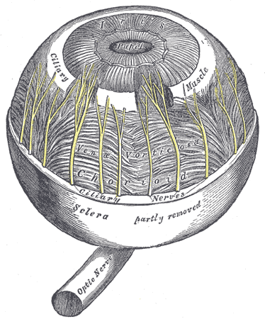
The choroid and iris.
-
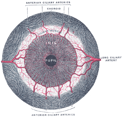
Iris, front view.
-
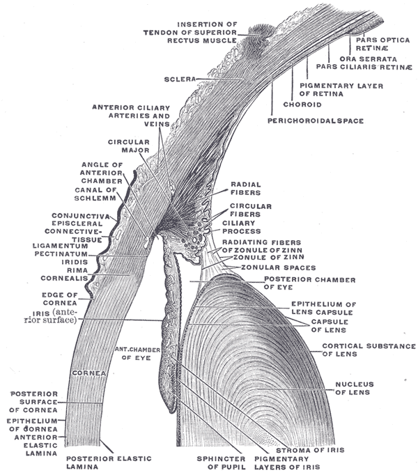
The upper half of a sagittal section through the front of the eyeball.


External links
- Histology image: 08010loa – Histology Learning System at Boston University
- Template:UMichAtlas - "Sagittal Section Through the Eyeball"
ar:قزحية ca:Iris (anatomia) da:Regnbuehinde de:Iris (Auge) el:Ίρις (οφθαλμού) eo:Iriso ko:홍채 it:Iride (anatomia) he:קשתית lt:Rainelė nl:Iris (anatomie) no:Regnbuehinne sk:Dúhovka (anatómia) sv:Regnbågshinna