Inferior cervical ganglion
Template:Infobox Nerve Editor-In-Chief: C. Michael Gibson, M.S., M.D. [1]
The inferior cervical ganglion is situated between the base of the transverse process of the last cervical vertebra and the neck of the first rib, on the medial side of the costocervical artery.
Its form is irregular; it is larger in size than the middle cervical ganglion, and is frequently fused with the first thoracic ganglion.
Path
It is probably formed by the coalescence of two ganglia which correspond to the seventh and eighth cervical nerves.
It is connected to the middle cervical ganglion by two or more cords, one of which forms a loop around the subclavian artery and supplies offsets to it. This loop is named the ansa subclavia (Vieussenii).
The ganglion sends gray rami communicantes to the seventh and eighth cervical nerves.
Branches
It gives off the inferior cardiac nerve, and offsets to bloodvessels.
Inferior cardiac nerve
Offsets to bloodvessels
The offsets to bloodvessels form plexuses on the subclavian artery and its branches.
The plexus on thevertebral artery is continued on to the basilar, posterior cerebral, and cerebellar arteries.
The plexus on the inferior thyroid artery accompanies the artery to the thyroid gland, and communicates with the recurrent and external laryngeal nerves, with the superior cardiac nerve, and with the plexus on the common carotid artery.
Additional images
-
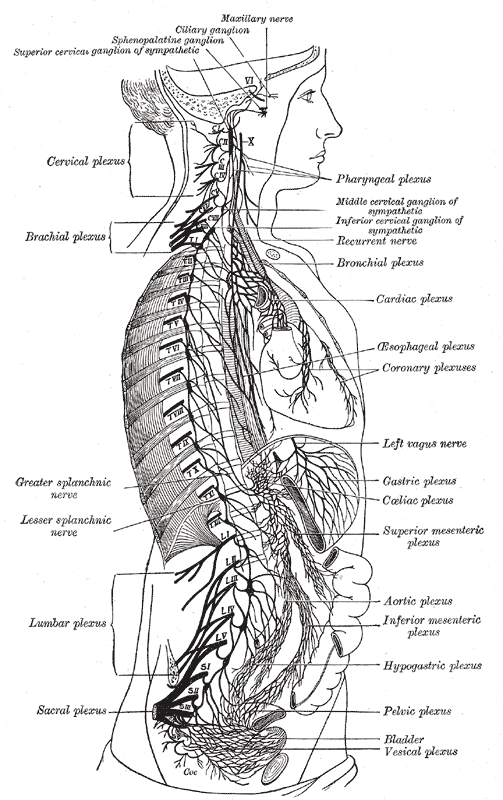 The right sympathetic chain and its connections with the thoracic, abdominal, and pelvic plexuses.

External links
- Template:GPnotebook
- Template:SUNYAnatomyLabs - "The Sympathetic Trunk and Cervical Ganglia"