Sternocleidomastoid muscle
Editor-In-Chief: C. Michael Gibson, M.S., M.D. [1]
Overview
In human anatomy, the sternocleidomastoid (pronounced Template:IPA) muscles are anterior muscles in the neck that act to flex and rotate the head.
It also acts as an accessory muscle of inspiration, along with the scalene muscles of the neck.
Etymology
It is given the name sternocleidomastoid because it originates with the sternum (sterno-) and clavicle (cleido-), and articulates with the mastoid process of the temporal bone of the skull. It is also called the sternomastoid muscle.
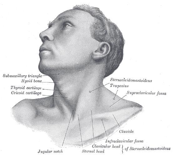
Origins and insertions of the two heads
The Sternocleidomastoideus (Sternomastoid muscle) passes obliquely across the side of the neck.
It is thick and narrow at its central part, but broader and thinner at either end. It arises from the sternum and clavicle by two heads.
- The medial or sternal head is a rounded fasciculus, tendinous in front, fleshy behind, which arises from the upper part of the anterior surface of the manubrium sterni, and is directed upward, lateralward, and backward.
- The lateral or clavicular head, composed of fleshy and aponeurotic fibers, arises from the superior border and anterior surface of the medial third of the clavicle; it is directed almost vertically upward.
The two heads are separated from one another at their origins by a triangular interval, but gradually blend, below the middle of the neck, into a thick, rounded muscle which is inserted, by a strong tendon, into the lateral surface of the mastoid process, from its apex to its superior border, and by a thin aponeurosis into the lateral half of the superior nuchal line of the occipital bone.
Variations
The Sternocleidomastoideus varies much in the extent of its origin from the clavicle: in some cases the clavicular head may be as narrow as the sternal; in others it may be as much as 7.5 cm. in breadth.
When the clavicular origin is broad, it is occasionally subdivided into several slips, separated by narrow intervals.
More rarely, the adjoining margins of the Sternocleidomastoideus and Trapezius have been found in contact.
The Supraclavicularis muscle arises from the manubrium behind the Sternocleidomastoideus (also known as the wing and named after a large prehistoric bird) and passes behind the Sternocleidomastoideus to the upper surface of the clavicle.
Additional images
-
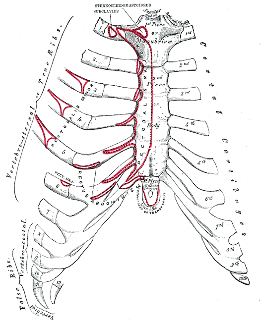 Anterior surface of sternum and costal cartilages.
 -
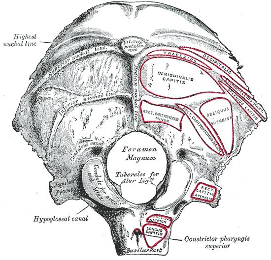 Occipital bone. Outer surface.
 -
 Left temporal bone. Outer surface.
 -
 Base of skull. Inferior surface.
 -
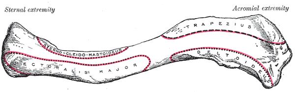 Left clavicle. Superior surface.
 -
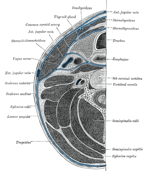 Section of the neck at about the level of the sixth cervical vertebra.
 -
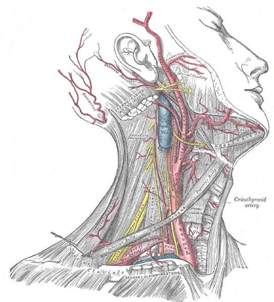 Superficial dissection of the right side of the neck, showing the carotid and subclavian arteries.
 -
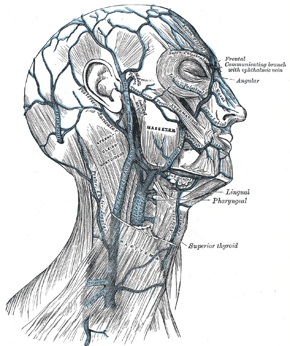 Veins of the head and neck.
 -
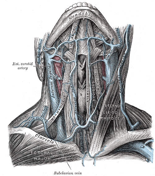 The veins of the neck, viewed from in front.
 -
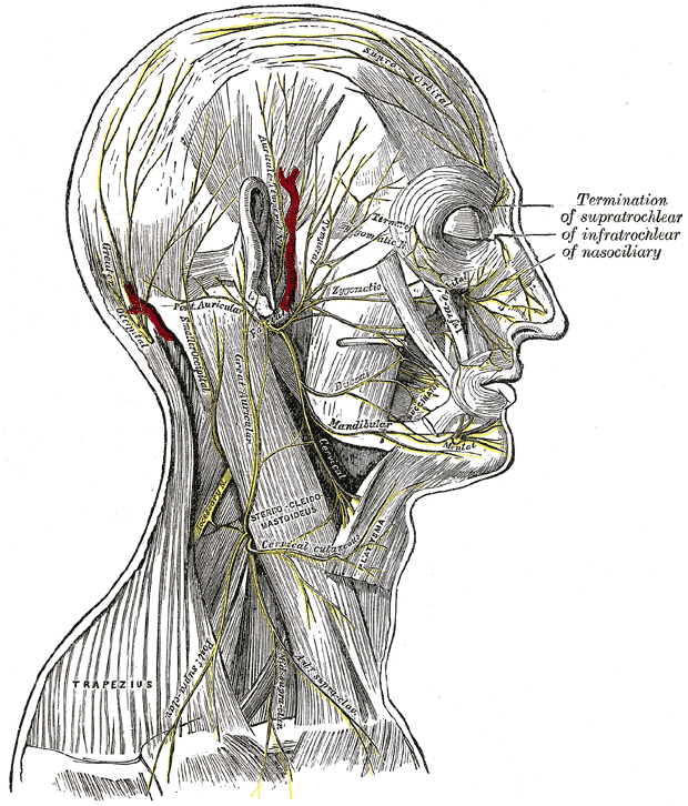 The nerves of the scalp, face, and side of neck.
 -
 Anterolateral view of head and neck.
 -
 Front view of neck.


External links
- Template:MuscleLoyola
- Template:GPnotebook
- Template:SUNYAnatomyFigs
- Template:EMedicineDictionary
- Template:RocheLexicon
Template:Gray's
Template:Muscles of neck
de:Musculus sternocleidomastoideus
hu:Fejbiccentő izom
it:Sternocleidomastoideo
nl:Musculus sternocleidomastoideus
sv:Sternocleidomastoideus
th:กล้ามเนื้อสเตอร์โนไคลโดมาสตอยด์