Otic ganglion
Template:Infobox Nerve Editor-In-Chief: C. Michael Gibson, M.S., M.D. [1]
The otic ganglion is a small, ovalshaped, flattened parasympathetic ganglion of a reddish-gray color, located immediately below the foramen ovale.
Location and relations
It is related to the maxillary and internal pterygoid nerves, to the cartilaginous part of the Eustachian tube, to the origin of the tensor palati muscle, and to the middle meningeal artery.
It surrounds the origin of the nerve to the Pterygoideus internus.
It is in relation, laterally, with the trunk of the mandibular nerve at the point where the motor and sensory roots join; medially, with the cartilaginous part of the auditory tube, and the origin of the Tensor veli palatini; posteriorly, with the middle meningeal artery.
Filaments
Functionally, it gives off filaments:
- posteriorly on the lateral surface of the Eustachian tube to the tensor tympani
- anteriorly to the tensor palati muscle
- to the inferior maxillary nerve, making up its motor root.
- to the auriculotemporal nerve, making up its sensory root.
Branches of Communication
It is connected by two or three short filaments with the nerve to the Pterygoideus internus, from which it may obtain a motor, and possibly a sensory root.
It communicates with the glossopharyngeal and facial nerves, through the lesser superficial petrosal nerve continued from the tympanic plexus, and through this nerve it probably receives a root from the glossopharyngeal and a motor root from the facial; its sympathetic root consists of a filament from the plexus surrounding the middle meningeal artery.
The fibers from the glossopharyngeal which pass to the otic ganglion in the small superficial petrosal are supposed to be parasympathetic efferent (preganglionic) fibers from the dorsal nucleus or inferior salivatory nucleus of the medulla.
Fibers (postganglionic) from the otic ganglion with which these form synapses are supposed to pass with the auriculotemporal nerve to the parotid gland.
A slender filament (sphenoidal) ascends from it to the nerve of the Pterygoid canal, and a small branch connects it with the chorda tympani.
Distribution
Its branches of distribution are: a filament to the Tensor tympani, and one to the Tensor veli palatini.
The former passes backward, lateral to the auditory tube; the latter arises from the ganglion, near the origin of the nerve to the Pterygoideus internus, and is directed forward.
The fibers of these nerves are, however, mainly derived from the nerve to the Pterygoideus internus.
Additional images
-
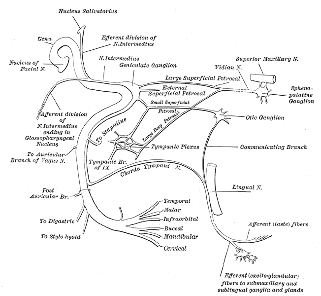 Plan of the facial and intermediate nerves and their communication with other nerves.
 -
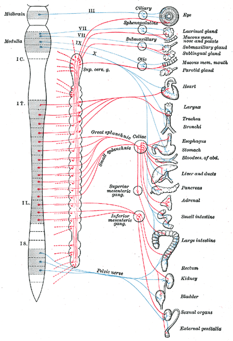 Diagram of efferent sympathetic nervous system.


References
- Shimizu T., "Distribution and pathway of the cerebrovascular nerve fibers from the otic ganglion in the rat: anterograde tracing study."; J Auton Nerv Syst. 1994 Sep;49(1):47-54. [2]
External links
- Template:NormanAnatomy (Template:NormanAnatomyFig, Template:NormanAnatomyFig)
- Template:EMedicineDictionary
Template:Gray's
Template:Trigeminal nerve
Template:Autonomic