Maxillary nerve
Template:Infobox Nerve Editor-In-Chief: C. Michael Gibson, M.S., M.D. [1]
The maxillary nerve (superior maxillary nerve), or second division of the trigeminal, is a sensory nerve.
It is intermediate, both in position and size, between the ophthalmic nerve and the mandibular nerve.
Path
Anterior to the trigeminal ganglion, the maxillary nerve passes through the cavernous sinus and exits the skull through the foramen rotundum.
It begins at the middle of the trigeminal ganglion as a flattened plexiform band, and, passing horizontally forward, it leaves the skull through the foramen rotundum, where it becomes more cylindrical in form, and firmer in texture.
It then crosses the pterygopalatine fossa, inclines lateralward on the back of the maxilla, and enters the orbit through the inferior orbital fissure; it traverses the infraorbital groove and canal in the floor of the orbit, and appears upon the face at the infraorbital foramen.
At its termination, the nerve lies beneath the quadratus labii superioris, and divides into a leash of branches which spread out upon the side of the nose, the lower eyelid, and the upper lip, joining with filaments of the facial nerve.
Branches
Its branches may be divided into four groups, according as they are given off in the cranium, in the pterygopalatine fossa, in the infraorbital canal, or on the face.
In the cranium
In the pterygopalatine fossa
- Zygomatic nerve (zygomaticotemporal nerve, zygomaticofacial nerve)
- Two small branches to the pterygopalatine ganglion (sometimes called the sphenopalatine ganglion)
- Posterior superior alveolar nerve
In the infraorbital canal
On the face
Additional images
-
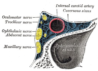
Oblique section through the cavernous sinus.
-
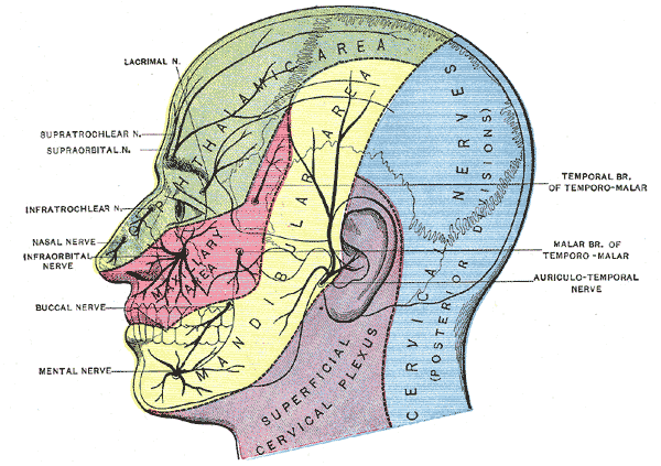
Dermatome distribution of the trigeminal nerve
-
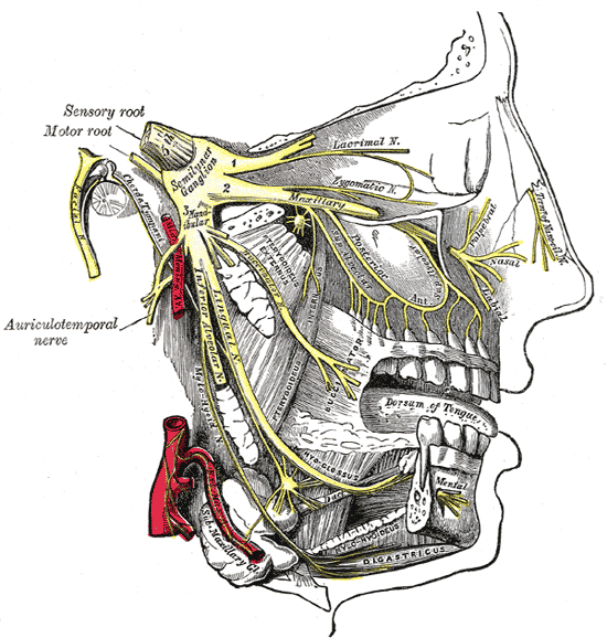
Distribution of the maxillary and mandibular nerves, and the submaxillary ganglion.
-
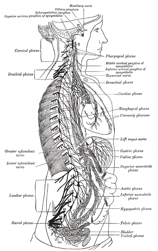
The right sympathetic chain and its connections with the thoracic, abdominal, and pelvic plexuses.


External links
de:Nervus maxillaris nl:Nervus maxillaris no:Nervus maxillaris