Hemorrhagic stroke CT
|
Hemorrhagic stroke Microchapters |
|
Diagnosis |
|---|
|
Treatment |
|
AHA/ASA Guidelines for the Management of Spontaneous Intracerebral Hemorrhage (2015) |
|
AHA/ASA Guidelines for the Management of Aneurysmal Subarachnoid Hemorrhage (2012) |
|
AHA/ASA Guideline Recommendation for the Primary Prevention of Stroke (2014) |
|
AHA/ASA Guideline Recommendations for Prevention of Stroke in Women (2014) Sex-Specific Risk Factors
Risk Factors Commoner in Women |
|
Case Studies |
|
Hemorrhagic stroke CT On the Web |
|
American Roentgen Ray Society Images of Hemorrhagic stroke CT |
Editor-In-Chief: C. Michael Gibson, M.S., M.D. [1]; Associate Editor(s)-in-Chief: Sara Mehrsefat, M.D. [2]
Overview
CT is very sensitive for identifying acute hemorrhage and is considered the gold standard.[1][2]
CT
CT is very sensitive for identifying acute hemorrhage and is considered the gold standard.[1][2]
Images
The following are images associated with Hemorrhagic stroke.[3]
-
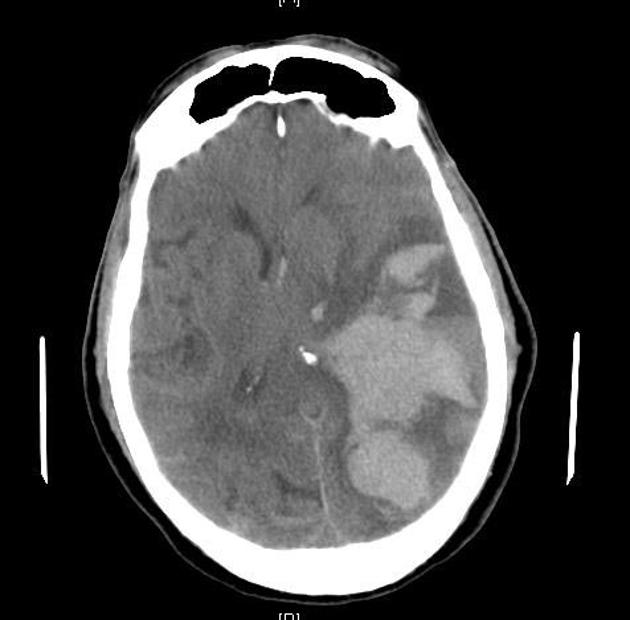
 Large hemorrhagic focus in the left cerebral hemisphere that extends to the infratentorial region with a significant mass effect
 -
 Large hemorrhagic focus in the left cerebral hemisphere that extends to the infratentorial region with a significant mass effect
 -
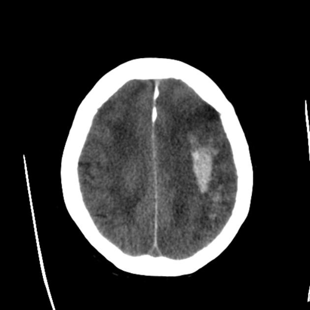
 Multiple descrete bilateral supratentorial intraparenchymal cerebral haemorrhage
 -
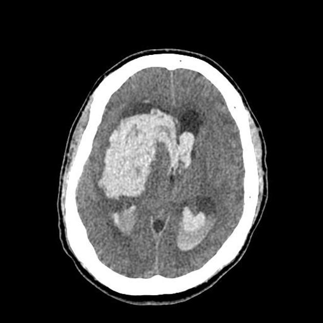
 Very large intracerebral haemorrhage on the left extends to involve the ventricles. It exerts marked mass effect
 -
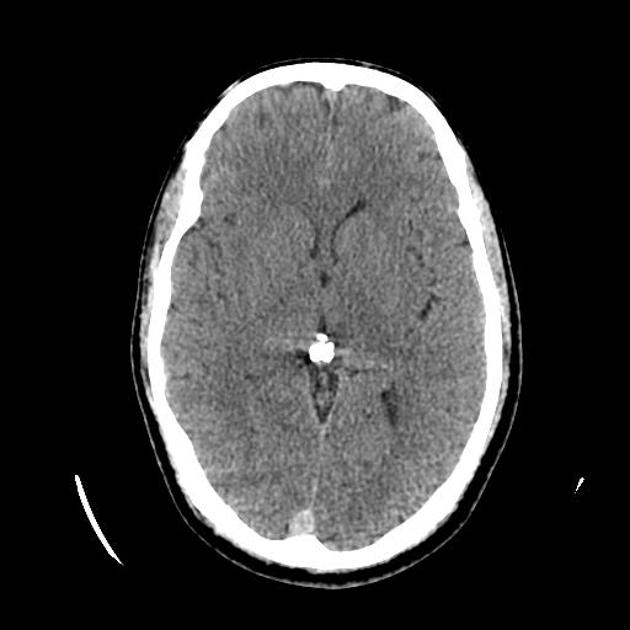
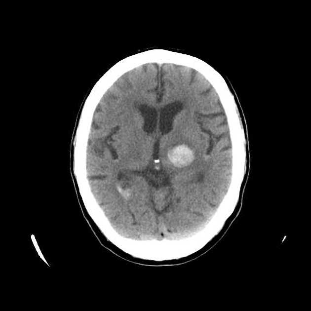 Hemorrhagic stroke in the left basal ganglia


References
- ↑ 1.0 1.1 Fiebach JB, Schellinger PD, Gass A, Kucinski T, Siebler M, Villringer A, Olkers P, Hirsch JG, Heiland S, Wilde P, Jansen O, Röther J, Hacke W, Sartor K; Kompetenznetzwerk Schlaganfall B5. Stroke magnetic resonance imaging is accurate in hyperacute intracerebral hemorrhage: a multicenter study on the validity of stroke imaging. Stroke. 2004;35:502– 506. doi: 10.1161/01.STR.0000114203.75678.8
- ↑ 2.0 2.1 Chalela JA, Kidwell CS, Nentwich LM, Luby M, Butman JA, Demchuk AM, Hill MD, Patronas N, Latour L, Warach S. Magnetic resonance imaging and computed tomography in emergency assessment of patients with suspected acute stroke: a prospective comparison. Lancet. 2007;369:293–298. doi: 10.1016/S0140-6736(07)60151-2.
- ↑ Intracerebral Hemotrrhage https://radiopaedia.org/cases/intracerebral-haemorrhage-2 Accessed on November 9, 2016