Medial longitudinal fasciculus
Overview
The medial longitudinal fasciculus (MLF) is a pair of crossed fiber tracts (group of axons), one on each side of the brainstem.
Function
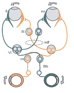
The MLF carries information about the direction that the eyes should move.
It yokes the cranial nerve nuclei III, IV and VI together, as well as the gaze centres and information about head movement (from cranial nerve VIII).
It also descends into the cervical spinal cord, and innervates some muscles of the neck.
Inputs
The MLF arises from the Vestibular nucleus (VN) and is thought to be involved in the maintenance of gaze. This is achieved by inputs to the VN from
- the Vestibulocochlear (8th cranial) nerve about head movements,
- gain adjustments from the flocculus of the cerebellum,
- head and neck propioceptors and foot and ankle muscle spindle, via the fastigial nucleus.
Pathology
Lesions of the MLF produce internuclear ophthalmoplegia. Lesions to the MLF are very common manifestations of the disease multiple sclerosis.where it most commonly presents as diplopia. These lesions cause damage to the ipsilateral (same side) eye.
History
In 1846 neurologist Benedict Stilling first referred to what is now known as the MLF as the acusticus, followed by Theodor Meynert in 1872 calling it posterior. But in 1891, Heinrich Schutz chose the name dorsal to describe the longitudinal bundle, "for brevity's sake". This name stuck despite other authors attempting further renaming (Ramon y Cajal's periependymal in 1904, Theodor Ziehen's nubecula dorsalis in 1913). But finally, it was Wilhelm His, Sr. who changed the name to medial for the sake of the Basle nomenclature to end the confusion.
Additional images
-
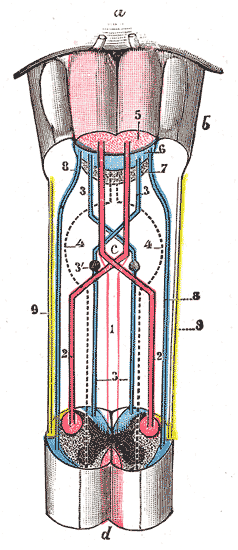 Decussation of pyramids.
 -
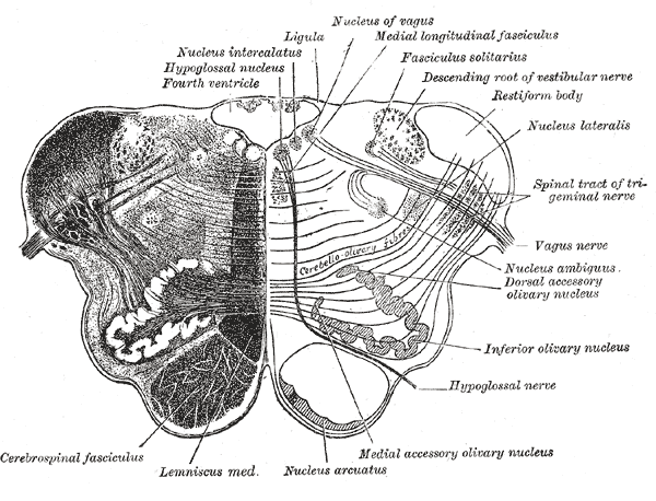 Transverse section of medulla oblongata below the middle of the olive.
 -


External links
- Template:EMedicineDictionary
- Template:UMichAtlas - "Brainstem, Cranial Nerve Nuclei, Sagittal Section, Medial View"
Template:Mesencephalon Template:Rhombencephalon Template:Eye-stub Template:Neuroscience-stub