Vestibulo-ocular reflex
Editor-In-Chief: C. Michael Gibson, M.S., M.D. [1]

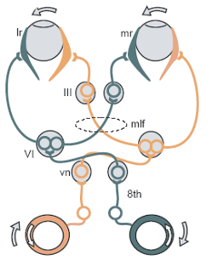
The green objects are excited, the orange ones inhibited.
The vestibulo-ocular reflex (VOR) or oculovestibular reflex is a reflex eye movement that stabilizes images on the retina during head movement by producing an eye movement in the direction opposite to head movement, thus preserving the image on the center of the visual field. For example, when the head moves to the right, the eyes move to the left, and vice versa. Since slight head movements are present all the time, the VOR is very important for stabilizing vision: patients whose VOR is impaired find it difficult to read using print, because they cannot stabilize the eyes during small head tremors. The VOR reflex does not depend on visual input and works even in total darkness or when the eyes are closed.
Gain
The "gain" of the VOR is defined as the change in the eye angle divided by the change in the head angle during the head turn. If the gain of the VOR is wrong (different than 1)—for example, if eye muscles are weak, or if a person puts on a new pair of eyeglasses—then head movements result in image motion on the retina, resulting in blurred vision. Under such conditions, motor learning adjusts the gain of the VOR to produce more accurate eye motion. This is what is referred to as VOR adaptation.
Ethanol consumption can disrupt the VOR, reducing dynamic visual acuity.[1]
Circuit
The main neural circuit for the horizontal VOR is fairly simple. Vestibular nuclei in the brainstem receive signals related to head movement from the Scarpa's ganglion located on CN VIII, or the vestibular nerve. From this Vestibular nuclei excitatory fibers cross to the contralateral CN VI nerve nucleus. There they synapse with 2 additional pathways. One projects directly to the lateral rectus of eye. Another nerve tract projects from the CN VI nucleus by the abducens internuclear interneurons or abducens interneurons to the oculomotor nuclei, which contain motorneurons that drive eye muscle activity, specifically activating the medial rectus muscles of the eye. Another pathway directly projects from the vestibular nucleus through the ascending tract of Dieters to the ipsilateral medial rectus motoneurons. In addition there are inhibitory vestibular pathways to the ipsilateral CN VI nucleus. However no direct vestibular neuron medial rectus motoneuron pathway exists. [2]
Role of cerebellum
The cerebellum is essential for motor learning to correct the VOR in order to ensure accurate eye movements. Motor learning in the VOR is in many ways analogous to classical eyeblink conditioning, since the circuits are homologous and the molecular mechanisms are similar.
See also
External links
- Motor Learning in the VOR in Mice at edboyden.org
- Review on VOR adaptation via slides at Johns Hopkins University
- Vestibulo-Ocular+Reflex at the US National Library of Medicine Medical Subject Headings (MeSH)
- ent/482 at eMedicine - "Vestibuloocular Reflex Testing"
References
- ↑ "Effect of Ethanol on visual-vestibular interactions during vertical linear body acceleration".
- ↑ Straka H, Dieringer N (2004). "Basic organization principles of the VOR: lessons from frogs". Prog. Neurobiol. 73 (4): 259–309. doi:10.1016/j.pneurobio.2004.05.003. PMID 15261395.