Inferior mesenteric artery
Editor-In-Chief: C. Michael Gibson, M.S., M.D. [1]
In human anatomy, the inferior mesenteric artery, often abbreviated as IMA, supplies the large intestine from the left colic (or splenic) flexure to the upper part of the rectum, which includes the descending colon, the sigmoid colon, and part of the rectum. Proximally, its territory of distribution overlaps (forms a watershed) with the middle colic artery, and therefore the superior mesenteric artery. The SMA and IMA anastomose via the marginal artery (artery of Drummond). The territory of distribution of the IMA is more or less equivalent to the embryonic hindgut.
Branching
The IMA branches off the anterior surface of the abdominal aorta below the renal artery branch points, and approximately midway between these and the aortic bifurcation (into the common iliac arteries).
The IMA has the following branches:
| Branch | notes |
| left colic artery | supplies descending colon |
| sigmoid branches | the most superior being described as 'the superior sigmoid artery' |
| superior rectal artery | effectively the terminal branch of the IMA (the continuation of the IMA after all other branches) |
All these arterial branches further divide into arcades which then supply the colon at regular intervals.
Associated veins
The IMA is accompanied along its course by a similarly named vein, the inferior mesenteric vein, which drains into the splenic vein.
The IMV therefore drains to the portal vein and does not fully mirror the course of the IMA.
Surgery and pathology
The IMA and/or its branches must be resected for a left hemicolectomy.
A horseshoe kidney, a rare (1 in 600) anomaly of the kidneys, will be positioned below the IMA.
Additional images
-
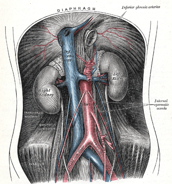 The abdominal aorta and its branches.
 -
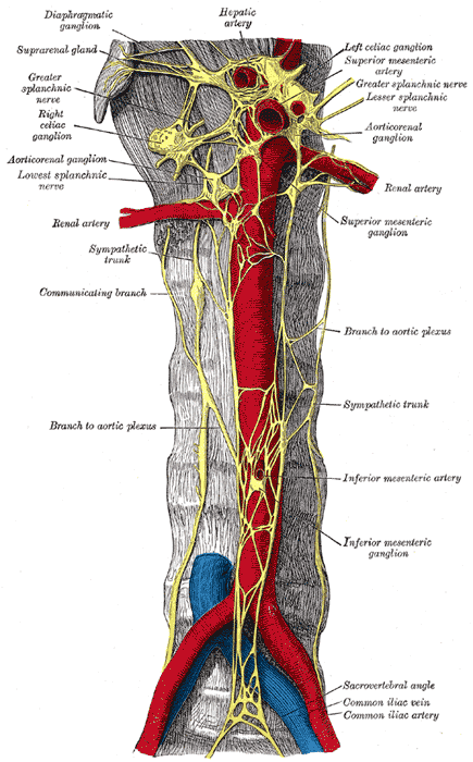 Abdominal portion of the sympathetic trunk, with the celiac plexus and hypogastric plexus.
 -
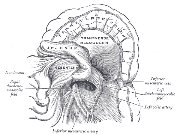 Duodenojejunal fossa.
 -
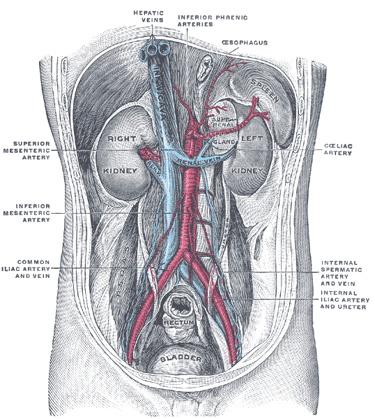 Posterior abdominal wall, after removal of the peritoneum, showing kidneys, suprarenal capsules, and great vessels.
 -
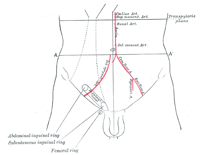 Front of abdomen, showing surface markings for arteries and inguinal canal.


External links
- Template:SUNYAnatomyFigs - "Branches of the inferior mesenteric artery."
- Template:SUNYAnatomyLabs - "Posterior Abdominal Wall: Branches of the Abdominal Aorta"
- Template:SUNYAnatomyImage
- Template:SUNYAnatomyImage
- Template:SUNYAnatomyImage
- Template:SUNYAnatomyImage
- Template:UMichAtlas - "Posterior Abdominal Wall, Dissection, Anterior View"
- Template:NormanAnatomy