Renal artery
|
WikiDoc Resources for Renal artery |
|
Articles |
|---|
|
Most recent articles on Renal artery Most cited articles on Renal artery |
|
Media |
|
Powerpoint slides on Renal artery |
|
Evidence Based Medicine |
|
Clinical Trials |
|
Ongoing Trials on Renal artery at Clinical Trials.gov Clinical Trials on Renal artery at Google
|
|
Guidelines / Policies / Govt |
|
US National Guidelines Clearinghouse on Renal artery
|
|
Books |
|
News |
|
Commentary |
|
Definitions |
|
Patient Resources / Community |
|
Patient resources on Renal artery Discussion groups on Renal artery Patient Handouts on Renal artery Directions to Hospitals Treating Renal artery Risk calculators and risk factors for Renal artery
|
|
Healthcare Provider Resources |
|
Causes & Risk Factors for Renal artery |
|
Continuing Medical Education (CME) |
|
International |
|
|
|
Business |
|
Experimental / Informatics |
Editor-In-Chief: C. Michael Gibson, M.S., M.D. [1]
The renal arteries normally arise off the side of the abdominal aorta, immediately below the superior mesenteric artery, and supply the kidneys with blood. Each is directed across the crus of the diaphragm, so as to form nearly a right angle with the aorta.
The renal arteries carry a large portion of total blood flow to the kidneys. Up to a third of total cardiac output can pass through the renal arteries to be filtered by the kidneys.
The arterial supply of the kidneys is variable and there may be one or more renal arteries supplying each kidney. It is located above the renal vein.
Asymmetries before reaching kidney
Due to the position of the aorta, the inferior vena cava and the kidneys in the body, the right renal artery is normally longer than the left renal artery.
- The right passes behind the inferior vena cava, the right renal vein, the head of the pancreas, and the descending part of the duodenum.
- The left is somewhat higher than the right; it lies behind the left renal vein, the body of the pancreas and the lienal vein, and is crossed by the inferior mesenteric vein.
At kidney
Before reaching the hilus of the kidney, each artery divides into four or five branches; the greater number of these lie between the renal vein and ureter, the vein being in front, the ureter behind, but one or more branches are usually situated behind the ureter.
Each vessel gives off some small inferior suprarenal branches to the suprarenal gland, the ureter, and the surrounding cellular tissue and muscles.
One or two accessory renal arteries are frequently found, more especially on the left side they usually arise from the aorta, and may come off above or below the main artery, the former being the more common position. Instead of entering the kidney at the hilus, they usually pierce the upper or lower part of the gland.
Diseases of the renal arteries
Renal artery stenosis, or narrowing of one or both renal arteries will lead to hypertension as the affected kidneys release renin to increase blood pressure to preserve perfusion to the kidneys. RAS is diagnosed with an MRA (magnet-resonance scan) of abdomen. It is treated with the use of balloon angioplasty and stents, if necessary.
Atherosclerosis can also affect the renal arteries and can lead to poor perfusion of the kidneys leading to reduced kidney function and, possibly, renal failure.
Additional images
-
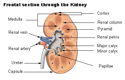 Frontal section through the kidney
 -
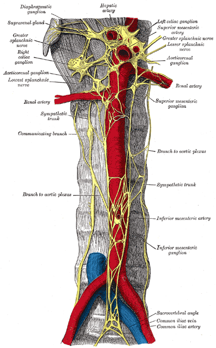 Abdominal portion of the sympathetic trunk, with the celiac and hypogastric plexuses.
 -
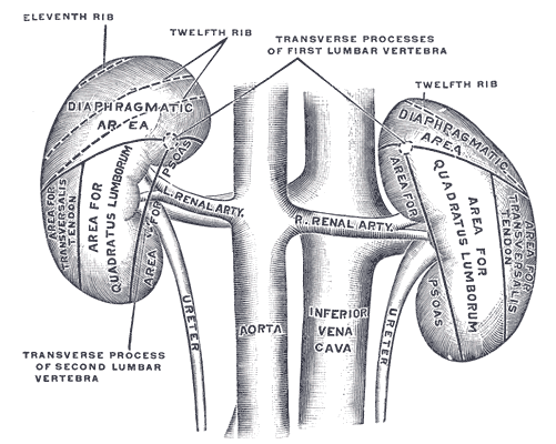 The posterior surfaces of the kidneys, showing areas of relation to the parietes.
 -
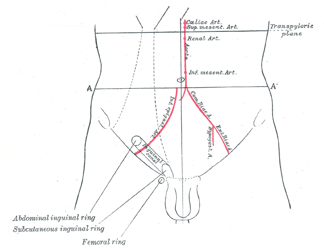 Front of abdomen, showing surface markings for arteries and inguinal canal.


External links
- Template:MedlinePlusImage
- Template:SUNYAnatomyLabs - "Posterior Abdominal Wall: Branches of the Abdominal Aorta"
Template:Arteries of thorax and abdomen Template:Kidney