Small intestine cancer pathophysiology: Difference between revisions
No edit summary |
No edit summary |
||
| Line 5: | Line 5: | ||
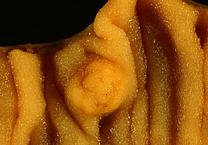
[[Adenocarcinoma]] is the most common sub-type of small intestine cancer. Second most common is [[carcinoid tumor]]. Adenocarcinomas can be polypoid, infiltrating or they appear as annular constricting lesions in small intestine. On gross pathology, napkin ring appearance or polypoidal fungatining mass are characteristic findings of small intestine cancer. Carcinoid tumors of the smalls intestine are mostly associated with malignant tumors of the other sites. Gastrointestinal stromal tumors (GISTs) are the most common benign tumors of the gastrointestinal (GI) tract. Small intestinal lymphomas are of low-grade histology and arise from mucosal-associated lymphoid tissues (MALT). | [[Adenocarcinoma]] is the most common sub-type of small intestine cancer. Second most common is [[carcinoid tumor]]. Adenocarcinomas can be polypoid, infiltrating or they appear as annular constricting lesions in small intestine. On gross pathology, napkin ring appearance or polypoidal fungatining mass are characteristic findings of small intestine cancer. Carcinoid tumors of the smalls intestine are mostly associated with malignant tumors of the other sites. Gastrointestinal stromal tumors (GISTs) are the most common benign tumors of the gastrointestinal (GI) tract. Small intestinal lymphomas are of low-grade histology and arise from mucosal-associated lymphoid tissues (MALT). | ||
==Pathophysiology== | |||
*Pathophysiology of the small intestinal cancers is not much studied domain, as it is a rare condition. | *Pathophysiology of the small intestinal cancers is not much studied domain, as it is a rare condition. | ||
| Line 18: | Line 18: | ||
**High concentration of lymphoid tissue | **High concentration of lymphoid tissue | ||
==Associations== | |||
Some GIT disorders such as inflammatory diseases of GIT may predispose to malignancy. Some of the associations are:<ref name="pmid11588539">{{cite journal |vauthors=Gill SS, Heuman DM, Mihas AA |title=Small intestinal neoplasms |journal=J. Clin. Gastroenterol. |volume=33 |issue=4 |pages=267–82 |date=October 2001 |pmid=11588539 |doi= |url=}}</ref> | Some GIT disorders such as inflammatory diseases of GIT may predispose to malignancy. Some of the associations are:<ref name="pmid11588539">{{cite journal |vauthors=Gill SS, Heuman DM, Mihas AA |title=Small intestinal neoplasms |journal=J. Clin. Gastroenterol. |volume=33 |issue=4 |pages=267–82 |date=October 2001 |pmid=11588539 |doi= |url=}}</ref> | ||
| Line 58: | Line 58: | ||
*Stromal tumors are most common in the stomach, 60–70% of the stromal tumors, followed by small intestine which makes 20–25% of all the stromal tumors of GI tract, colon and rectum makes 5% and esophagus is less than 5%. | *Stromal tumors are most common in the stomach, 60–70% of the stromal tumors, followed by small intestine which makes 20–25% of all the stromal tumors of GI tract, colon and rectum makes 5% and esophagus is less than 5%. | ||
==Genetics== | |||
Cancer of small intestine can arise sporadically or they are associated with genetic diseases. | Cancer of small intestine can arise sporadically or they are associated with genetic diseases. | ||
| Line 81: | Line 81: | ||
==Gross Pathology== | |||
| Line 103: | Line 103: | ||
==Microscopic Pathology== | |||
Revision as of 21:33, 11 January 2019
|
Small intestine cancer Microchapters |
|
Diagnosis |
|---|
|
Treatment |
|
Case Studies |
|
Small intestine cancer pathophysiology On the Web |
|
American Roentgen Ray Society Images of Small intestine cancer pathophysiology |
|
Risk calculators and risk factors for Small intestine cancer pathophysiology |
Editor-In-Chief: C. Michael Gibson, M.S., M.D. [1]; Associate Editor(s)-in-Chief: Qurrat-ul-ain Abid, M.D.[2], Parminder Dhingra, M.D. [3]
Overview
Adenocarcinoma is the most common sub-type of small intestine cancer. Second most common is carcinoid tumor. Adenocarcinomas can be polypoid, infiltrating or they appear as annular constricting lesions in small intestine. On gross pathology, napkin ring appearance or polypoidal fungatining mass are characteristic findings of small intestine cancer. Carcinoid tumors of the smalls intestine are mostly associated with malignant tumors of the other sites. Gastrointestinal stromal tumors (GISTs) are the most common benign tumors of the gastrointestinal (GI) tract. Small intestinal lymphomas are of low-grade histology and arise from mucosal-associated lymphoid tissues (MALT).
Pathophysiology
- Pathophysiology of the small intestinal cancers is not much studied domain, as it is a rare condition.
- Studies are being conducted to evaluate association with environmental risk factors.[1]
- Pathophysiology of small intestinal cancers depend on the histological subtype.
- Duodenal tumors are more common than the tumors of jejunum and illeum.[2]
- Adenomas arise more proximally in the duodenum and lymphomas arise in jejunum and ileum.[3]
- Low susceptibility of the small intestine to malignant changes can be explained by following:
- Short exposure of the mucosa to carcinogens due to rapid transit of contents.
- Liquid nature of the contents and less mucosal irritation.
- Low bacterial load
- High concentration of lymphoid tissue
Associations
Some GIT disorders such as inflammatory diseases of GIT may predispose to malignancy. Some of the associations are:[4]
- Familial Adenomatous Polyposis Coli: adenoma, adenocarcinoma
- Peutz-Jeghers syndrome: hamartomatous polyps
- Chronic intestinal inflammatory disorders e.g., Crohn's disease: adenocarcinoma
- Celiac sprue: lymphoma
- Von Recklinghausen's disease: paraganglioma
- Immunoproliferative small intestinal disease: small intestinal lymphoma
Adenocarcinoma:
- Adenocarcinoma of the smalls intestine originate locally or can be associated with malignant tumors of other sites.
- Rarely it can develop fom malignant changes in polyps present in the small intestine.
Carcinoid Tumors:
- Carcinoid tumors are the second most common cancer of the small intestine.
- It is a slow growing tumor of small intestine arising as a subset of neuroendocrine cells.
- Carcinoid tumors of the smalls intestine are mostly associated with malignant tumors of the other sites. [5]
- Small Intestinal neuroendocrine tumors (SI-NETs) are the most common gastrointestinal neuroendocrine tumors.[6]
- The oncofetal protein IMP3 is a marker that plays a role in the growth of neuroendocrine cells.[7]
Non-Hodgkin Lymphoma:
- After stomach, small intestine is the most common extra-nodal site of presentation of non-Hodgkin lymphomas and it represents 4% to 20% of all the non-Hodgkin lymphomas.
- Some of the association of non-Hodgkin lymphomas are :[8]
- Helicobacter pylori infection
- Immunosuppression after solid-organ transplantation
- Celiac disease
- Inflammatory bowel disease
- Human immunodeficiency virus (HIV)
Small intestinal stromal tumors (GISTs):
- GISTs are the most benign tumors of GIT and rarely can be malignant.
- They typically develop in older age.
- Stromal tumors are most common in the stomach, 60–70% of the stromal tumors, followed by small intestine which makes 20–25% of all the stromal tumors of GI tract, colon and rectum makes 5% and esophagus is less than 5%.
Genetics
Cancer of small intestine can arise sporadically or they are associated with genetic diseases.
Adenocarcinoma:
- Small intestine adenocarcinomas arise from adenomas.
- Adenoma-carcinoma sequence is thought to play the role in development of small intestine adenocarcinoma but is not understood by the researchers yet.[9]
- Adenocarcinoma is thought to be associated with some genetic diseases such as:
- Herditary Non-Polyposis colosis(HNPCC)[10][11]
- Gardner's syndrome[12]
- Peutz-Jeghers syndrome[13]
- Familial Adenosis Polyposis (FAP)[14]
Dietary factors, tobacco, and obesity
Carcinoid Tumors:
Non-Hodgkin Lymphoma:
Small intestinal stromal tumors (GISTs):
Gross Pathology
Adenocarcinoma:
- Adenocarcinomas can be polypoid, infiltrating, or as annular constricting lesions is small intestine. Polyps and adenomas of small intestine are considered precursor lesions of adenocarcinoma.[15]

Carcinoid Tumors:
- They are diverse neoplasms emerging from the endocrine cells of the intestinal mucosa.[7]


Non-Hodgkin Lymphoma:
Small intestinal stromal tumors (GISTs):
- Instestinal stromal cell tumors should be differentiated from leiomyosarcomas and leiomyomas. They differ clinically and pathogenetically from leiomyosarcomas and leiomyomas. Leimyomas occur in the GI tract, commonly in the esophagus.

Microscopic Pathology
Adenocarcinoma:
- Primary adenocarcinoma consists of 40% of cases of malignant tumors of small intestine and it is the most common histologic type. [5]
Carcinoid Tumors:
- These tumors originate from enterochromaffin (EC) cells and secrete serotonin.[16]

Non-Hodgkin Lymphoma:
- Small intestinal lymphomas are of low-grade histology and arise from mucosal-associated lymphoid tissues (MALT) present in ileum and jejunum.Sometimies distinct clinicopathologic entities arise from these mucosal-associated lymphoid tissues (MALT), such as immunoproliferative small intestinal disease, primary intestinal T-cell lymphoma and multiple lymphomatous polyposis.[8]
Small intestinal stromal tumors (GISTs):
- One of the subset of intestinal stromal cell tumors is the GI autonomic nerve tumors (GANTs). Stromal tumors can be differentiated from other tumors of small intestine by their cell specific markers.
- GISTs express following stromal cell markers:[17]
- 70% of GISTs are positive for CD34
- 20–30% are positive for smooth muscle actin (SMA)
- 10% are positive for S100 protein
- <5% are positive for desmin
References
- ↑ Severson RK, Schenk M, Gurney JG, Weiss LK, Demers RY (February 1996). "Increasing incidence of adenocarcinomas and carcinoid tumors of the small intestine in adults". Cancer Epidemiol. Biomarkers Prev. 5 (2): 81–4. PMID 8850266.
- ↑ Dabaja BS, Suki D, Pro B, Bonnen M, Ajani J (August 2004). "Adenocarcinoma of the small bowel: presentation, prognostic factors, and outcome of 217 patients". Cancer. 101 (3): 518–26. doi:10.1002/cncr.20404. PMID 15274064.
- ↑ Chow JS, Chen CC, Ahsan H, Neugut AI (August 1996). "A population-based study of the incidence of malignant small bowel tumours: SEER, 1973-1990". Int J Epidemiol. 25 (4): 722–8. PMID 8921448.
- ↑ Gill SS, Heuman DM, Mihas AA (October 2001). "Small intestinal neoplasms". J. Clin. Gastroenterol. 33 (4): 267–82. PMID 11588539.
- ↑ 5.0 5.1 Barclay TH, Schapira DV (March 1983). "Malignant tumors of the small intestine". Cancer. 51 (5): 878–81. PMID 6821853.
- ↑ Modlin, Irvin M.; Champaneria, Manish C.; Chan, Anthony K.C.; Kidd, Mark (2007). "A Three-Decade Analysis of 3,911 Small Intestinal Neuroendocrine Tumors: The Rapid Pace of No Progress". The American Journal of Gastroenterology. 102 (7): 1464–1473. doi:10.1111/j.1572-0241.2007.01185.x. ISSN 0002-9270.
- ↑ 7.0 7.1 Massironi S, Del Gobbo A, Cavalcoli F, Fiori S, Conte D, Pellegrinelli A, Milione M, Ferrero S (November 2017). "IMP3 expression in small-intestine neuroendocrine neoplasms: a new predictor of recurrence". Endocrine. 58 (2): 360–367. doi:10.1007/s12020-017-1249-x. PMID 28210937.
- ↑ 8.0 8.1 Crump M, Gospodarowicz M, Shepherd FA (June 1999). "Lymphoma of the gastrointestinal tract". Semin. Oncol. 26 (3): 324–37. PMID 10375089.
- ↑ Wheeler JM, Warren BF, Mortensen NJ, Kim HC, Biddolph SC, Elia G, Beck NE, Williams GT, Shepherd NA, Bateman AC, Bodmer WF (February 2002). "An insight into the genetic pathway of adenocarcinoma of the small intestine". Gut. 50 (2): 218–23. PMC 1773117. PMID 11788563.
- ↑ Zhang MQ, Chen ZM, Wang HL (April 2006). "Immunohistochemical investigation of tumorigenic pathways in small intestinal adenocarcinoma: a comparison with colorectal adenocarcinoma". Mod. Pathol. 19 (4): 573–80. doi:10.1038/modpathol.3800566. PMID 16501564.
- ↑ Rodriguez-Bigas MA, Vasen HF, Lynch HT, Watson P, Myrhøj T, Järvinen HJ, Mecklin JP, Macrae F, St John DJ, Bertario L, Fidalgo P, Madlensky L, Rozen P (July 1998). "Characteristics of small bowel carcinoma in hereditary nonpolyposis colorectal carcinoma. International Collaborative Group on HNPCC". Cancer. 83 (2): 240–4. PMID 9669805.
- ↑ Kawashima A, Goldman SM, Fishman EK, Kuhlman JE, Onitsuka H, Fukuya T, Masuda K (February 1994). "CT of intraabdominal desmoid tumors: is the tumor different in patients with Gardner's disease?". AJR Am J Roentgenol. 162 (2): 339–42. doi:10.2214/ajr.162.2.8310922. PMID 8310922.
- ↑ Giardiello FM, Brensinger JD, Tersmette AC, Goodman SN, Petersen GM, Booker SV, Cruz-Correa M, Offerhaus JA (December 2000). "Very high risk of cancer in familial Peutz-Jeghers syndrome". Gastroenterology. 119 (6): 1447–53. PMID 11113065.
- ↑ Abrahams NA, Halverson A, Fazio VW, Rybicki LA, Goldblum JR (November 2002). "Adenocarcinoma of the small bowel: a study of 37 cases with emphasis on histologic prognostic factors". Dis. Colon Rectum. 45 (11): 1496–502. doi:10.1097/01.DCR.0000034134.49346.5E. PMID 12432298.
- ↑ Levine JS, Ahnen DJ (December 2006). "Clinical practice. Adenomatous polyps of the colon". N. Engl. J. Med. 355 (24): 2551–7. doi:10.1056/NEJMcp063038. PMID 17167138.
- ↑ Sei Y, Feng J, Zhao X, Forbes J, Tang D, Nagashima K, Hanson J, Quezado MM, Hughes MS, Wank SA (July 2016). "Polyclonal Crypt Genesis and Development of Familial Small Intestinal Neuroendocrine Tumors". Gastroenterology. 151 (1): 140–51. doi:10.1053/j.gastro.2016.03.007. PMC 5578471. PMID 27003604.
- ↑ Schlotzhauer WS, Chortyk OT, Austin PR (1976). "Pyrolysis of chitin, a potential tobacco extender". J Agric Food Chem. 24 (1): 177–80. PMID 1432-2307 Check
|pmid=value (help).