Pneumoconiosis diagnostic criteria: Difference between revisions
| (39 intermediate revisions by the same user not shown) | |||
| Line 3: | Line 3: | ||
{{CMG}}; {{AE}} [[User:Dushka|Dushka Riaz, MD]] | {{CMG}}; {{AE}} [[User:Dushka|Dushka Riaz, MD]] | ||
== Overview == | ==Overview== | ||
The initial imaging done for [[pneumoconiosis]] is a [[Chest X-ray|chest x-ray]]. This serves as a [[screening test]]. [[High-resolution CT]] follows and is more [[Sensitivity (test)|sensitive]] and [[Specificity|specific]]. [[HRCT]] can identify those [[diseases]] missed by [[chest radiograph]]. [[Pathognomonic]] for [[asbestosis]] is pleural thickening with pleural [[plaques]]. [[Silicosis]] would show round opacities in the upper [[lung]]. Massive [[fibrosis]] can be seen in both [[coal worker's pneumoconiosis]] and [[silicosis]]. <ref name="pmid1410306">{{cite journal| author=Remy-Jardin M, Remy J, Farre I, Marquette CH| title=Computed tomographic evaluation of silicosis and coal workers' pneumoconiosis. | journal=Radiol Clin North Am | year= 1992 | volume= 30 | issue= 6 | pages= 1155-76 | pmid=1410306 | doi= | pmc= | url=https://www.ncbi.nlm.nih.gov/entrez/eutils/elink.fcgi?dbfrom=pubmed&tool=sumsearch.org/cite&retmode=ref&cmd=prlinks&id=1410306 }} </ref> <ref name="pmid1987601">{{cite journal| author=Akira M, Yokoyama K, Yamamoto S, Higashihara T, Morinaga K, Kita N | display-authors=etal| title=Early asbestosis: evaluation with high-resolution CT. | journal=Radiology | year= 1991 | volume= 178 | issue= 2 | pages= 409-16 | pmid=1987601 | doi=10.1148/radiology.178.2.1987601 | pmc= | url=https://www.ncbi.nlm.nih.gov/entrez/eutils/elink.fcgi?dbfrom=pubmed&tool=sumsearch.org/cite&retmode=ref&cmd=prlinks&id=1987601 }} </ref> <ref name="pmid14576443">{{cite journal| author=Copley SJ, Wells AU, Sivakumaran P, Rubens MB, Lee YC, Desai SR | display-authors=etal| title=Asbestosis and idiopathic pulmonary fibrosis: comparison of thin-section CT features. | journal=Radiology | year= 2003 | volume= 229 | issue= 3 | pages= 731-6 | pmid=14576443 | doi=10.1148/radiol.2293020668 | pmc= | url=https://www.ncbi.nlm.nih.gov/entrez/eutils/elink.fcgi?dbfrom=pubmed&tool=sumsearch.org/cite&retmode=ref&cmd=prlinks&id=14576443 }} </ref> <ref name="pmid33153681">{{cite journal| author=Walkoff L, Hobbs S| title=Chest Imaging in the Diagnosis of Occupational Lung Diseases. | journal=Clin Chest Med | year= 2020 | volume= 41 | issue= 4 | pages= 581-603 | pmid=33153681 | doi=10.1016/j.ccm.2020.08.007 | pmc= | url=https://www.ncbi.nlm.nih.gov/entrez/eutils/elink.fcgi?dbfrom=pubmed&tool=sumsearch.org/cite&retmode=ref&cmd=prlinks&id=33153681 }} </ref> | |||
== Diagnostic Study of Choice == | ==Diagnostic Study of Choice== | ||
=== Study of choice === | ===Study of choice=== | ||
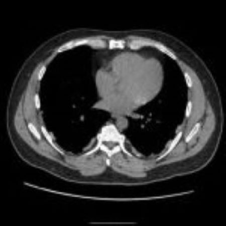
[[Image:Asbestosis-5.jpg|thumb|200px|left|Asbestosis with pleural plaques - Case courtesy of Dr Hani Makky Al Salam, <a href="https://radiopaedia.org/">Radiopaedia.org</a>. From the case <a href="https://radiopaedia.org/cases/45002">rID: 45002</a>]] | |||
1. Radiologic tests must be performed to test for [[asbestosis]] when: | |||
*The patient has had exposure to [[asbestos]] (with Helsinki criteria indicating the dose being at least 25 fibre/ml.years) | |||
* | *The [[CT scan]] would show [[pulmonary fibrosis]], pleural thickening and pleural plaques. <ref name="pmid23034792">{{cite journal| author=Darnton A, Hodgson J, Benson P, Coggon D| title=Mortality from asbestosis and mesothelioma in Britain by birth cohort. | journal=Occup Med (Lond) | year= 2012 | volume= 62 | issue= 7 | pages= 549-52 | pmid=23034792 | doi=10.1093/occmed/kqs119 | pmc=3471357 | url=https://www.ncbi.nlm.nih.gov/entrez/eutils/elink.fcgi?dbfrom=pubmed&tool=sumsearch.org/cite&retmode=ref&cmd=prlinks&id=23034792 }} </ref> <ref name="pmid9322824">{{cite journal| author=| title=Asbestos, asbestosis, and cancer: the Helsinki criteria for diagnosis and attribution. | journal=Scand J Work Environ Health | year= 1997 | volume= 23 | issue= 4 | pages= 311-6 | pmid=9322824 | doi= | pmc= | url=https://www.ncbi.nlm.nih.gov/entrez/eutils/elink.fcgi?dbfrom=pubmed&tool=sumsearch.org/cite&retmode=ref&cmd=prlinks&id=9322824 }}</ref> | ||
* | |||
2. The best test for [[silicosis]] is a [[High Resolution CT|high resolution CT]]: | |||
The | |||
=== Diagnostic Criteria === | *It would show widespread [[fibrosis]] with bilateral [[nodules]] and evidence of involvement of [[lymph nodes]]. It can be confirmed with lung biopsy showing [[acellular]] whorls, and bi-refringent crystals of [[silica]]. <ref name="pmid23708110">{{cite journal| author=Cullinan P, Reid P| title=Pneumoconiosis. | journal=Prim Care Respir J | year= 2013 | volume= 22 | issue= 2 | pages= 249-52 | pmid=23708110 | doi=10.4104/pcrj.2013.00055 | pmc=6442808 | url=https://www.ncbi.nlm.nih.gov/entrez/eutils/elink.fcgi?dbfrom=pubmed&tool=sumsearch.org/cite&retmode=ref&cmd=prlinks&id=23708110 }} </ref> | ||
3. [[Coal worker's pneumoconiosis]] also presents similarly to [[silicosis]] on [[HRCT]]: | |||
*[[Nodular]] opacities are seen with preference for upper lobes as well as massive [[pulmonary fibrosis]]. <ref name="pmid2217770">{{cite journal| author=Remy-Jardin M, Degreef JM, Beuscart R, Voisin C, Remy J| title=Coal worker's pneumoconiosis: CT assessment in exposed workers and correlation with radiographic findings. | journal=Radiology | year= 1990 | volume= 177 | issue= 2 | pages= 363-71 | pmid=2217770 | doi=10.1148/radiology.177.2.2217770 | pmc= | url=https://www.ncbi.nlm.nih.gov/entrez/eutils/elink.fcgi?dbfrom=pubmed&tool=sumsearch.org/cite&retmode=ref&cmd=prlinks&id=2217770 }} </ref> | |||
4. [[Berylliosis]] cases should have testing completed as well: | |||
*A blood beryllium lymphocyte proliferation test is the screening test of choice for those suspected of [[berylliosis]]. <ref name="pmid25398119">{{cite journal| author=Balmes JR, Abraham JL, Dweik RA, Fireman E, Fontenot AP, Maier LA | display-authors=etal| title=An official American Thoracic Society statement: diagnosis and management of beryllium sensitivity and chronic beryllium disease. | journal=Am J Respir Crit Care Med | year= 2014 | volume= 190 | issue= 10 | pages= e34-59 | pmid=25398119 | doi=10.1164/rccm.201409-1722ST | pmc= | url=https://www.ncbi.nlm.nih.gov/entrez/eutils/elink.fcgi?dbfrom=pubmed&tool=sumsearch.org/cite&retmode=ref&cmd=prlinks&id=25398119 }} </ref> | |||
*CT scans would show [[Ground glass opacification on CT|ground glass opacities]] as the earliest sign. <ref name="pmid12362066">{{cite journal| author=Maier LA| title=Clinical approach to chronic beryllium disease and other nonpneumoconiotic interstitial lung diseases. | journal=J Thorac Imaging | year= 2002 | volume= 17 | issue= 4 | pages= 273-84 | pmid=12362066 | doi=10.1097/00005382-200210000-00004 | pmc= | url=https://www.ncbi.nlm.nih.gov/entrez/eutils/elink.fcgi?dbfrom=pubmed&tool=sumsearch.org/cite&retmode=ref&cmd=prlinks&id=12362066 }} </ref> <ref name="pmid21084914">{{cite journal| author=Sharma N, Patel J, Mohammed TL| title=Chronic beryllium disease: computed tomographic findings. | journal=J Comput Assist Tomogr | year= 2010 | volume= 34 | issue= 6 | pages= 945-8 | pmid=21084914 | doi=10.1097/RCT.0b013e3181ef214e | pmc= | url=https://www.ncbi.nlm.nih.gov/entrez/eutils/elink.fcgi?dbfrom=pubmed&tool=sumsearch.org/cite&retmode=ref&cmd=prlinks&id=21084914 }} </ref> | |||
===Diagnostic Criteria=== | |||
To be qualified as a [[pneumoconiosis]] or [[occupational disease]] there must be four criteria met: | |||
*This includes documented exposure to the particle. | *This includes documented exposure to the particle. | ||
*[[Latent period]] before the development of [[symptoms]]. | *[[Latent period]] before the development of [[symptoms]]. | ||
Latest revision as of 23:00, 26 April 2021
|
Pneumoconiosis Microchapters |
|
Diagnosis |
|---|
|
Treatment |
|
Case Studies |
|
Pneumoconiosis diagnostic criteria On the Web |
|
American Roentgen Ray Society Images of Pneumoconiosis diagnostic criteria |
|
Risk calculators and risk factors for Pneumoconiosis diagnostic criteria |
Editor-In-Chief: C. Michael Gibson, M.S., M.D. [1]; Associate Editor(s)-in-Chief: Dushka Riaz, MD
Overview
The initial imaging done for pneumoconiosis is a chest x-ray. This serves as a screening test. High-resolution CT follows and is more sensitive and specific. HRCT can identify those diseases missed by chest radiograph. Pathognomonic for asbestosis is pleural thickening with pleural plaques. Silicosis would show round opacities in the upper lung. Massive fibrosis can be seen in both coal worker's pneumoconiosis and silicosis. [1] [2] [3] [4]
Diagnostic Study of Choice
Study of choice

1. Radiologic tests must be performed to test for asbestosis when:
- The patient has had exposure to asbestos (with Helsinki criteria indicating the dose being at least 25 fibre/ml.years)
- The CT scan would show pulmonary fibrosis, pleural thickening and pleural plaques. [5] [6]
2. The best test for silicosis is a high resolution CT:
- It would show widespread fibrosis with bilateral nodules and evidence of involvement of lymph nodes. It can be confirmed with lung biopsy showing acellular whorls, and bi-refringent crystals of silica. [7]
3. Coal worker's pneumoconiosis also presents similarly to silicosis on HRCT:
- Nodular opacities are seen with preference for upper lobes as well as massive pulmonary fibrosis. [8]
4. Berylliosis cases should have testing completed as well:
- A blood beryllium lymphocyte proliferation test is the screening test of choice for those suspected of berylliosis. [9]
- CT scans would show ground glass opacities as the earliest sign. [10] [11]
Diagnostic Criteria
To be qualified as a pneumoconiosis or occupational disease there must be four criteria met:
- This includes documented exposure to the particle.
- Latent period before the development of symptoms.
- Clinical signs and symptoms that entail the disease
- Exclusion of other disease modalities. [12]
References
- ↑ Remy-Jardin M, Remy J, Farre I, Marquette CH (1992). "Computed tomographic evaluation of silicosis and coal workers' pneumoconiosis". Radiol Clin North Am. 30 (6): 1155–76. PMID 1410306.
- ↑ Akira M, Yokoyama K, Yamamoto S, Higashihara T, Morinaga K, Kita N; et al. (1991). "Early asbestosis: evaluation with high-resolution CT". Radiology. 178 (2): 409–16. doi:10.1148/radiology.178.2.1987601. PMID 1987601.
- ↑ Copley SJ, Wells AU, Sivakumaran P, Rubens MB, Lee YC, Desai SR; et al. (2003). "Asbestosis and idiopathic pulmonary fibrosis: comparison of thin-section CT features". Radiology. 229 (3): 731–6. doi:10.1148/radiol.2293020668. PMID 14576443.
- ↑ Walkoff L, Hobbs S (2020). "Chest Imaging in the Diagnosis of Occupational Lung Diseases". Clin Chest Med. 41 (4): 581–603. doi:10.1016/j.ccm.2020.08.007. PMID 33153681 Check
|pmid=value (help). - ↑ Darnton A, Hodgson J, Benson P, Coggon D (2012). "Mortality from asbestosis and mesothelioma in Britain by birth cohort". Occup Med (Lond). 62 (7): 549–52. doi:10.1093/occmed/kqs119. PMC 3471357. PMID 23034792.
- ↑ "Asbestos, asbestosis, and cancer: the Helsinki criteria for diagnosis and attribution". Scand J Work Environ Health. 23 (4): 311–6. 1997. PMID 9322824.
- ↑ Cullinan P, Reid P (2013). "Pneumoconiosis". Prim Care Respir J. 22 (2): 249–52. doi:10.4104/pcrj.2013.00055. PMC 6442808. PMID 23708110.
- ↑ Remy-Jardin M, Degreef JM, Beuscart R, Voisin C, Remy J (1990). "Coal worker's pneumoconiosis: CT assessment in exposed workers and correlation with radiographic findings". Radiology. 177 (2): 363–71. doi:10.1148/radiology.177.2.2217770. PMID 2217770.
- ↑ Balmes JR, Abraham JL, Dweik RA, Fireman E, Fontenot AP, Maier LA; et al. (2014). "An official American Thoracic Society statement: diagnosis and management of beryllium sensitivity and chronic beryllium disease". Am J Respir Crit Care Med. 190 (10): e34–59. doi:10.1164/rccm.201409-1722ST. PMID 25398119.
- ↑ Maier LA (2002). "Clinical approach to chronic beryllium disease and other nonpneumoconiotic interstitial lung diseases". J Thorac Imaging. 17 (4): 273–84. doi:10.1097/00005382-200210000-00004. PMID 12362066.
- ↑ Sharma N, Patel J, Mohammed TL (2010). "Chronic beryllium disease: computed tomographic findings". J Comput Assist Tomogr. 34 (6): 945–8. doi:10.1097/RCT.0b013e3181ef214e. PMID 21084914.
- ↑ Epler GR (1992). "Clinical overview of occupational lung disease". Radiol Clin North Am. 30 (6): 1121–33. PMID 1410303.