Paget's disease of the breast pathophysiology: Difference between revisions
(Mahshid) |
Preeti Singh (talk | contribs) No edit summary |
||
| Line 3: | Line 3: | ||
{{CMG}};{{AE}} {{PSK}} | {{CMG}};{{AE}} {{PSK}} | ||
==Overview== | ==Overview== | ||
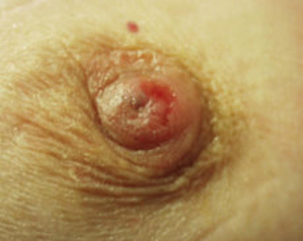
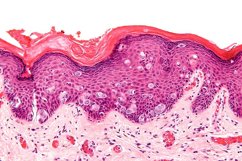
On gross pathology, eczematoid, [[Erythema|erythematous]], moist or crusted lesion, with or without fine scaling, infiltration of the [[nipple]], and inversion of the nipple are characteristic findings of Paget's disease of the [[breast]]. | On gross pathology, eczematoid, [[Erythema|erythematous]], moist or crusted lesion, with or without fine scaling, infiltration of the [[nipple]], and inversion of the nipple are characteristic findings of Paget's disease of the [[breast]]. [[Eczematoid|Eczema]] changes of the nipple-areolar complex are said to occur due to invasion of the overlying epidermis by malignant (Paget) cells. On microscopic histopathological analysis, epidermal Paget cells which are malignant glandular [[epithelial cells]] organized in groups with nest-like patterns or gland-like structures and are preferably located in the epidermal basal layer are characteristic findings of Paget's disease of the breast. | ||
==Pathophysiology== | ==Pathophysiology== | ||
===Pathogenesis=== | ===Pathogenesis=== | ||
The pathogenesis of Paget’s disease of the breast still remains controversial and supported by two different theories:<ref name="SubramanianBirch2007">{{cite journal|last1=Subramanian|first1=Ashok|last2=Birch|first2=Hilary|last3=McAvinchey|first3=Rita|last4=Stacey-Clear|first4=Adam|title=Pagets disease of uncertain origin: case report|journal=International Seminars in Surgical Oncology|volume=4|issue=1|year=2007|pages=12|issn=14777800|doi=10.1186/1477-7800-4-12}}</ref> | The pathogenesis of Paget’s disease of the breast still remains controversial and supported by two different theories:<ref name="SubramanianBirch2007">{{cite journal|last1=Subramanian|first1=Ashok|last2=Birch|first2=Hilary|last3=McAvinchey|first3=Rita|last4=Stacey-Clear|first4=Adam|title=Pagets disease of uncertain origin: case report|journal=International Seminars in Surgical Oncology|volume=4|issue=1|year=2007|pages=12|issn=14777800|doi=10.1186/1477-7800-4-12}}</ref><ref name="Lopes FilhoLopes2015">{{cite journal|last1=Lopes Filho|first1=Lauro Lourival|last2=Lopes|first2=Ione Maria Ribeiro Soares|last3=Lopes|first3=Lauro Rodolpho Soares|last4=Enokihara|first4=Milvia M. S. S.|last5=Michalany|first5=Alexandre Osores|last6=Matsunaga|first6=Nobuo|title=Mammary and extramammary Paget's disease|journal=Anais Brasileiros de Dermatologia|volume=90|issue=2|year=2015|pages=225–231|issn=1806-4841|doi=10.1590/abd1806-4841.20153189}}</ref> | ||
*Epidermotropic theory | *Epidermotropic theory | ||
*Intraepidermal transformation theory | *Intraepidermal transformation theory | ||
Revision as of 21:12, 3 March 2019
|
Paget's disease of the breast Microchapters |
|
Differentiating Paget's disease of the breast from other Diseases |
|---|
|
Diagnosis |
|
Treatment |
|
Case Studies |
|
Paget's disease of the breast pathophysiology On the Web |
|
American Roentgen Ray Society Images of Paget's disease of the breast pathophysiology |
|
Directions to Hospitals Treating Paget's disease of the breast |
|
Risk calculators and risk factors for Paget's disease of the breast pathophysiology |
Editor-In-Chief: C. Michael Gibson, M.S., M.D. [4];Associate Editor(s)-in-Chief: Suveenkrishna Pothuru, M.B,B.S. [5]
Overview
On gross pathology, eczematoid, erythematous, moist or crusted lesion, with or without fine scaling, infiltration of the nipple, and inversion of the nipple are characteristic findings of Paget's disease of the breast. Eczema changes of the nipple-areolar complex are said to occur due to invasion of the overlying epidermis by malignant (Paget) cells. On microscopic histopathological analysis, epidermal Paget cells which are malignant glandular epithelial cells organized in groups with nest-like patterns or gland-like structures and are preferably located in the epidermal basal layer are characteristic findings of Paget's disease of the breast.
Pathophysiology
Pathogenesis
The pathogenesis of Paget’s disease of the breast still remains controversial and supported by two different theories:[1][2]
- Epidermotropic theory
- Intraepidermal transformation theory
Epidermotropic Theory
- It is thought that malignant epithelial cells from intraductal carcinoma, extend into the overlying epidermis through mammary duct epithelium and proliferate in the epidermis causing thickening of the nipple and areolar skin.[1]
- Normal epidermal keratinocytes produce and release the mobility factor heregulin-alpha which is chemotactic for heregulin receptors (Her-2) and coreceptors Her 3 and Her 4 which are produced by Pagets cells. This is thought to result in migration of these cells to the nipple epidermis.
- This is supported by the observation that Paget cells often share cell surface markers with the underlying breast carcinoma (e.g CAM 5.2, CEA, c-erb 2 and EMA).
Intraepidermal transformation theory
- The degeneration of preexisting cells, considers that Paget cells are keratinocytes that have undergone malignant transformation and that the disease is an in situ carcinoma regardless of the underlying intraductal carcinoma. The underlying intraductal carcinoma is simply coexisting with this disease.
Gross Pathology
- On gross pathology, eczematoid, erythematous, moist or crusted lesion, with or without fine scaling, infiltration of the nipple, and inversion of the nipple are characteristic findings of Paget's disease of the breast.[2]
 |
Microscopic pathology
- Paget's disease of the breast is histopathologically characterized by epidermal Paget cells, which are malignant glandular epithelial cells with abundant and clear cytoplasm, usually containing mucin and pleomorphic and hyperchromatic nucleus.[2]
- These cells appear organized in groups, with nest-like patterns or gland-like structures, and are preferably located in the epidermal basal layer.
- The number of cells varies from a few to large quantities; even completely replacing the epidermal cells.
- Invasion of adnexal structures can occur.
- Orthokeratosis and parakeratosis may be present.
- The dermis displays reactive characteristics, with telangiectasia, chronic inflammation, and ulceration in more advanced cases.
- There are several histologic variants of Paget's disease include:[3]
- Adenocarcinoma-like cell type
- Spindle cell type
- Anaplastic cell type
- Acantholytic cell type
- Pigmented cell type
 |
Immunohistochemistry
- Immunohistochemistry is very useful in Paget's disease of the breast for differential diagnoses and histogenesis.[2]
- Overexpression of the low molecular weight cytokeratins, notably CK7, and lack of expression of high molecular weight cytokeratins, such as CK10, CK14 and CK20 are observed.
- Paget cells have the same immunohistochemical staining pattern as the underlying breast cancer cells.
- Furthermore, they also express carcinoembryonic antigen, epithelial membrane antigen, and some mucins.
- Since breast cancers associated to Paget's disease are poorly differentiated, estrogen and progesterone antigens are frequently negative.
- Mori et al found overexpression of oncogenic ras and p21 in mammary and extramammary diseases.
- Paget cells express p53, p21, Ki-67, cyclin D1, androgen receptors and Her-2 oncoprotein.
References
- ↑ 1.0 1.1 Subramanian, Ashok; Birch, Hilary; McAvinchey, Rita; Stacey-Clear, Adam (2007). "Pagets disease of uncertain origin: case report". International Seminars in Surgical Oncology. 4 (1): 12. doi:10.1186/1477-7800-4-12. ISSN 1477-7800.
- ↑ 2.0 2.1 2.2 2.3 Lopes Filho, Lauro Lourival; Lopes, Ione Maria Ribeiro Soares; Lopes, Lauro Rodolpho Soares; Enokihara, Milvia M. S. S.; Michalany, Alexandre Osores; Matsunaga, Nobuo (2015). "Mammary and extramammary Paget's disease". Anais Brasileiros de Dermatologia. 90 (2): 225–231. doi:10.1590/abd1806-4841.20153189. ISSN 1806-4841.
- ↑ 3.0 3.1 Image courtesy of Dr Garth Kruger. Radiopaedia (original file [1]). [http://radiopaedia.org/licence Creative Commons BY-SA-NC
- ↑ Paget's disease of the breast. [2] (original file [3])