Lisch nodules are well defined [[Melanoma|melanocytic]] [[hamartomas]] of the [[iris]]. Lisch nodules generally [[Appearance|appear]] as dome-shaped gelatinous [[Mass|masses]]. Lisch nodules masses are more commonly develop on the surface of the [[iris]], also known as [[iris]] [[hamartomas]]. Lisch nodules are gold-tan to brown in [[color]], they may [[Growth|grow]] up to 2 mm in [[diameter]] and can be of different sizes on the same [[iris]]. Lisch nodules arise from [[mast cells]], pigmented [[cells]] and [[fibroblast]]-like [[cells]]. The presence of Lisch nodules is the most common [[clinical]] [[Medical sign|sign]] of [[Neurofibromatosis 1]]; ninety-three [[Percentage|percent]] of cases are [[Bilateral|bilaterally]] affected and an [[average]] of 25 [[nodules]] can be counted on each [[iris]]. Once [[iris]] [[hamartomas]] have [[Development|developed]], they remain stable throughout [[life]]. In 80% of [[eyes]], Lisch nodules may be found in the inferior [[Quadrant|quadrants]] of the [[iris]] and this may be related to greater [[sun exposure]], one of the postulated factors in the [[development]] of these [[benign]] tumefactions.
==Historical Perspective==
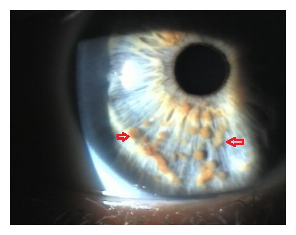
*[[File:Lisch Nodule.jpg|alt=Lisch nodules|thumb|Multiple small, oval, yellow-brown [[papules]] (Lisch nodules) in the right [[Iris (anatomy)|iris]](Red arrows). case courtesy by E. G. Adams et al.<ref>{{Cite web|url=https://www.ncbi.nlm.nih.gov/pmc/articles/PMC3350217/|title=Multiple, Unilateral Lisch Nodules in the Absence of Other Manifestations of Neurofibromatosis Type 1|last=|first=|date=|website=|archive-url=|archive-date=|dead-url=|access-date=}}</ref>]]In 1882, [[Neurofibromatosis 1|Neurofibromatosis]] ([[NF]]), described by Friedrich Daniel [[Von Recklinghausen neurofibromatosis|Von Recklinghausen]].<ref name="pmid23793209">{{cite journal| author=Antônio JR, Goloni-Bertollo EM, Trídico LA| title=Neurofibromatosis: chronological history and current issues. | journal=An Bras Dermatol | year= 2013 | volume= 88 | issue= 3 | pages= 329-43 | pmid=23793209 | doi=10.1590/abd1806-4841.20132125 | pmc=3754363 | url=https://www.ncbi.nlm.nih.gov/entrez/eutils/elink.fcgi?dbfrom=pubmed&tool=sumsearch.org/cite&retmode=ref&cmd=prlinks&id=23793209 }}</ref><ref name="Hosoi1931">{{cite journal|last1=Hosoi|first1=Kiyoshi|title=MULTIPLE NEUROFIBROMATOSIS (von RECKLINGHAUSEN'S DISEASE)|journal=Archives of Surgery|volume=22|issue=2|year=1931|pages=258|issn=0272-5533|doi=10.1001/archsurg.1931.01160020081004}}</ref>
*[[NF]] is a neuroectodermal abnormality constituted by a [[set]] of [[clinical]] [[symptoms]] that compromise the [[skin]], [[nervous system]], [[bones]], [[eyes]] and other sites.
*Lisch Nodules were named after the infamous Austrian [[ophthalmology|ophthalmologist]] Karl Lisch(1907-1999) who was also called as "Ophthalmological Pope".
*Besides general [[ophthalmology]], Lisch was interested in [[scientific]] [[research]]. He published more than 120 [[scientific]] papers in German [[Science (journal)|science]] journals.
*In 1937, Karl Lisch published an article on the [[iris]] [[hamartomas]] and their association with [[neurofibromatosis 1]], now known as "Lisch nodules", while at the University Eye Clinic in Munich.
*Lisch'a article described his [[Observation|observations]] in 3 [[patients]] with [[neurofibromatosis]]:<ref name="pmid20418991">{{cite journal| author=Gabhane SK, Kotwal MN, Bobhate SK| title=Segmental neurofibromatosis: a report of 3 cases. | journal=Indian J Dermatol | year= 2010 | volume= 55 | issue= 1 | pages= 105-8 | pmid=20418991 | doi=10.4103/0019-5154.60366 | pmc=2856359 | url=https://www.ncbi.nlm.nih.gov/entrez/eutils/elink.fcgi?dbfrom=pubmed&tool=sumsearch.org/cite&retmode=ref&cmd=prlinks&id=20418991 }}</ref><ref name="Dimitrova2009">{{cite journal|last1=Dimitrova|first1=Valentina|title=A CASE OF NEUROFIBROMATOSIS TYPE 1|journal=Journal of IMAB - Annual Proceeding (Scientific Papers)|volume=14, 1|issue=2008|year=2009|pages=63–67|issn=1312773X|doi=10.5272/jimab.14-1-2010.63}}</ref>
**[[Patient]] 1: A 39 [[year]] [[Old age|old]] [[male]] who had been affected with several [[nodules]] and [[Pigmented lesions|pigmented]] [[lesions]] on the [[skin]], typical of [[neurofibromatosis]], since the [[age]] of 15 [[Year|years]]. His mother and his sisters had a similar [[disorder]]. Lisch observed several [[brown]] [[nodules]] on the surface of the [[iris]]. The [[nodules]] could be seen even without the [[slit lamp]] due to the greyish-blue [[color]] of the [[iris]].
**[[Patient]] 2: 27 [[year]] [[Old age|old]] [[patient]] with similar [[cutaneous]] and [[iris]] [[lesions]] could be detected along with a [[family]] [[History and Physical examination|history]] of [[neurofibromatosis]]. In comparison to the first [[patient]] the [[iris]] [[nodules]] were much more pigmented.
**[[Patient]] 3: A 44 [[year]] [[Old age|old]] [[male]] suffered form [[bilateral]] [[optic]] [[nerve]] [[gliomas]] with [[Chiasma|chiasmal]] involvement. The [[slit lamp]] [[examination]] revealed tiny [[iris]] [[nodules]] in both [[eyes]].
==Classification==
* There is no established system for the classification of Lisch nodules.
==Pathophysiology==
*A [[hamartoma]] is defined as a [[benign]] [[tumor]] or [[nodular]] [[growth]] that is composed of [[Proliferation|proliferating]] [[Maturation|mature]] [[histologically]] normal [[cells]] that normally reside at the affected [[tissue]].<ref name="pmid19650418">{{cite journal| author=Terzi YK, Oguzkan-Balci S, Anlar B, Aysun S, Guran S, Ayter S| title=Reproductive decisions after prenatal diagnosis in neurofibromatosis type 1: importance of genetic counseling. | journal=Genet Couns | year= 2009 | volume= 20 | issue= 2 | pages= 195-202 | pmid=19650418 | doi= | pmc= | url=https://www.ncbi.nlm.nih.gov/entrez/eutils/elink.fcgi?dbfrom=pubmed&tool=sumsearch.org/cite&retmode=ref&cmd=prlinks&id=19650418 }}</ref><ref name="pmid19516012">{{cite journal| author=Boley S, Sloan JL, Pemov A, Stewart DR| title=A quantitative assessment of the burden and distribution of Lisch nodules in adults with neurofibromatosis type 1. | journal=Invest Ophthalmol Vis Sci | year= 2009 | volume= 50 | issue= 11 | pages= 5035-43 | pmid=19516012 | doi=10.1167/iovs.09-3650 | pmc=2883270 | url=https://www.ncbi.nlm.nih.gov/entrez/eutils/elink.fcgi?dbfrom=pubmed&tool=sumsearch.org/cite&retmode=ref&cmd=prlinks&id=19516012 }}</ref><ref name="pmid19539839">{{cite journal| author=Boyd KP, Korf BR, Theos A| title=Neurofibromatosis type 1. | journal=J Am Acad Dermatol | year= 2009 | volume= 61 | issue= 1 | pages= 1-14; quiz 15-6 | pmid=19539839 | doi=10.1016/j.jaad.2008.12.051 | pmc=2716546 | url=https://www.ncbi.nlm.nih.gov/entrez/eutils/elink.fcgi?dbfrom=pubmed&tool=sumsearch.org/cite&retmode=ref&cmd=prlinks&id=19539839 }}</ref><ref name="TheosKorf2006">{{cite journal|last1=Theos|first1=Amy|last2=Korf|first2=Bruce R.|title=Pathophysiology of Neurofibromatosis Type 1|journal=Annals of Internal Medicine|volume=144|issue=11|year=2006|pages=842|issn=0003-4819|doi=10.7326/0003-4819-144-11-200606060-00010}}</ref>
*[[NF1]] is due to [[mutations]] in the [[NF1]] [[gene]], located at [[chromosome]] 17q11.2.<ref name="pmid20422842">{{cite journal| author=Cohen R, Shuper A| title=[Developmental manifestation in children with neurofibromatosis type 1]. | journal=Harefuah | year= 2010 | volume= 149 | issue= 1 | pages= 49-52, 61 | pmid=20422842 | doi= | pmc= | url=https://www.ncbi.nlm.nih.gov/entrez/eutils/elink.fcgi?dbfrom=pubmed&tool=sumsearch.org/cite&retmode=ref&cmd=prlinks&id=20422842 }}</ref><ref name="pmid115721742">{{cite journal| author=de Goede-Bolder A, Cnossen MH, Dooijes D, van den Ouweland AM, Niermeijer MF| title=[From gene to disease; neurofibromatosis type 1]. | journal=Ned Tijdschr Geneeskd | year= 2001 | volume= 145 | issue= 36 | pages= 1736-8 | pmid=11572174 | doi= | pmc= | url=https://www.ncbi.nlm.nih.gov/entrez/eutils/elink.fcgi?dbfrom=pubmed&tool=sumsearch.org/cite&retmode=ref&cmd=prlinks&id=11572174 }}</ref>
*Lisch nodules are generally not present in central NF([[neurofibromatosis]])/NF([[neurofibromatosis]])-2.
*[[Neurofibromin]], the [[protein]] product encoded by the [[gene]], is expressed in many [[tissues]], including [[brain]], [[kidney]], [[spleen]], and [[thymus]].
*[[Mutations]] in the [[NF1]] [[gene]] result in loss of production or reduced [[Function (biology)|function]] of [[protein]]; this causes a wide spectrum of clinical findings, including [[NF1]]-associated [[Tumor|tumors]].<ref name="LubsBauer1991">{{cite journal|last1=Lubs|first1=Marie-Louise E.|last2=Bauer|first2=Mislen S.|last3=Formas|first3=Maria E.|last4=Djokic|first4=Borivoje|title=Lisch Nodules in Neurofibromatosis Type 1|journal=New England Journal of Medicine|volume=324|issue=18|year=1991|pages=1264–1266|issn=0028-4793|doi=10.1056/NEJM199105023241807}}</ref><ref name="Dimitrova20092">{{cite journal|last1=Dimitrova|first1=Valentina|title=A CASE OF NEUROFIBROMATOSIS TYPE 1|journal=Journal of IMAB - Annual Proceeding (Scientific Papers)|volume=14, 1|issue=2008|year=2009|pages=63–67|issn=1312773X|doi=10.5272/jimab.14-1-2010.63}}</ref>
*[[Histopathology|Histopathologically]], Lisch nodules are composed of [[melanocytes]] and [[spindle cells]], usually concentrated on the superficial layers of the [[iris]] [[stroma]].<ref name="pmid28979620">{{cite journal| author=Abaloun Y, Ajhoun Y| title=[Lisch nodule in neurofibromatosis type 1]. | journal=Pan Afr Med J | year= 2017 | volume= 27 | issue= | pages= 218 | pmid=28979620 | doi=10.11604/pamj.2017.27.218.11517 | pmc=5622834 | url=https://www.ncbi.nlm.nih.gov/entrez/eutils/elink.fcgi?dbfrom=pubmed&tool=sumsearch.org/cite&retmode=ref&cmd=prlinks&id=28979620 }}</ref><ref name="pmid150961513">{{cite journal| author=Richetta A, Giustini S, Recupero SM, Pezza M, Carlomagno V, Amoruso G et al.| title=Lisch nodules of the iris in neurofibromatosis type 1. | journal=J Eur Acad Dermatol Venereol | year= 2004 | volume= 18 | issue= 3 | pages= 342-4 | pmid=15096151 | doi=10.1111/j.1468-3083.2004.00915.x | pmc= | url=https://www.ncbi.nlm.nih.gov/entrez/eutils/elink.fcgi?dbfrom=pubmed&tool=sumsearch.org/cite&retmode=ref&cmd=prlinks&id=15096151 }}</ref><ref name="RichettaGiustini2004">{{cite journal|last1=Richetta|first1=A|last2=Giustini|first2=S|last3=Recupero|first3=SM|last4=Pezza|first4=M|last5=Carlomagno|first5=V|last6=Amoruso|first6=G|last7=Calvieri|first7=S|title=Lisch nodules of the iris in neurofibromatosis type 1|journal=Journal of the European Academy of Dermatology and Venereology|volume=18|issue=3|year=2004|pages=342–344|issn=0926-9959|doi=10.1111/j.1468-3083.2004.00915.x}}</ref>
*The [[spindle cells]] are larger than the normal [[Iris (anatomy)|iris]] [[melanocytes]].
*[[Immunohistochemical]] studies show positive reaction against the following
**[[Vimentin]]
**[[Smooth muscle]] [[actin]]
**[[Neuron]] specific [[enolase]].
{{SK}} Sakurai-lisch nodule
== Causes ==
* Lisch Nodules commonly associated with [[neurofibromatosis]] and is caused by [[Genetics|genetic]] defects or [[mutations]] that either are passed on by a parent or occur spontaneously at [[conception]].
==[[Lisch nodule overview|Overview]]==
==Differential diagnosis==
Lisch nodules are well defined melanocytic hamartomas of the iris. They appear as dome-shaped gelatinous masses developing on the surface of the iris. Also known as iris hamartomas, lisch nodules are gold-tan to brown in color, they may grow up to 2 mm in diameter and can be of different sizes on the same iris. Lisch nodules arise from mast cells, pigmented cells and fibroblast-like cells. The presence of Lisch nodules is the most common clinical sign of Neurofibromatosis 1; ninety-three percent of cases are bilaterally affected and an average of 25 nodules can be counted on each iris. Once iris hamartomas have developed, they remain stable throughout life. In 80% of eyes, Lisch nodules may be found in the inferior quadrants of the iris and this may be related to greater sun exposure, one of the postulated factors in the development of these benign tumefactions.
* Lisch nodules must be differentiated from [[Iris (anatomy)|Iris]] mammillations, [[Iridocorneal endothelial syndrome]], Rieger's anomaly or syndrome, [[Iris (anatomy)|Iris]] [[nevi]], [[Melanoma]], [[Inflammatory]] conditions such as [[Sarcoidosis]], [[Leprosy]], [[Tuberculosis]] and [[Syphilis]].<ref name="pmid6992584">{{cite journal| author=Radius RL, Herschler J| title=Histopathology in the iris-nevus (Cogan-Reese) syndrome. | journal=Am J Ophthalmol | year= 1980 | volume= 89 | issue= 6 | pages= 780-6 | pmid=6992584 | doi=10.1016/0002-9394(80)90165-8 | pmc= | url=https://www.ncbi.nlm.nih.gov/entrez/eutils/elink.fcgi?dbfrom=pubmed&tool=sumsearch.org/cite&retmode=ref&cmd=prlinks&id=6992584 }}</ref><ref name="pmid3190109">{{cite journal| author=Makley TA, Kapetansky FM| title=Iris nevus syndrome. | journal=Ann Ophthalmol | year= 1988 | volume= 20 | issue= 8 | pages= 311-5 | pmid=3190109 | doi= | pmc= | url=https://www.ncbi.nlm.nih.gov/entrez/eutils/elink.fcgi?dbfrom=pubmed&tool=sumsearch.org/cite&retmode=ref&cmd=prlinks&id=3190109 }}</ref>
==Historical Perspective==
== Epidemiology and Demographics ==
*Neurofibromatosis (NF), a disease described in 1882 by Friedrich Daniel Von Recklinghausen. <ref name="pmid23793209">{{cite journal| author=Antônio JR, Goloni-Bertollo EM, Trídico LA| title=Neurofibromatosis: chronological history and current issues. | journal=An Bras Dermatol | year= 2013 | volume= 88 | issue= 3 | pages= 329-43 | pmid=23793209 | doi=10.1590/abd1806-4841.20132125 | pmc=3754363 | url=https://www.ncbi.nlm.nih.gov/entrez/eutils/elink.fcgi?dbfrom=pubmed&tool=sumsearch.org/cite&retmode=ref&cmd=prlinks&id=23793209 }} </ref>
*NF is a neuroectodermal abnormality constituted by a set of clinical symptoms that compromise the skin, nervous system, bones, eyes and other sites.
*Lisch Nodules were named after the infamous Austrian [[ophthalmology|ophthalmologist]] Karl Lisch (1907-1999). <ref name="Singh2009">{{cite journal|last1=Singh|first1=Arun D.|title=Karl Lisch, MD: Remembered July 24, 1907 - February 5, 1999|journal=Ophthalmic Genetics|volume=21|issue=2|year=2009|pages=129–131|issn=1381-6810|doi=10.1076/1381-6810(200006)2121-8FT129}}</ref>
*Lisch was a well known ophthalmologist; his patients came from all parts of Austria, Germany, and Italy. In the region of North Tyrol he was called "Ophthalmological Pope".
*Besides general ophthalmology, Lisch was interested in scientific research. He published more than 120 scientific papers in German science journals.
*In 1937, Karl Lisch published an article on the iris hamartomas and their association with neurofibromatosis 1, now known as "Lisch nodules", while at the University Eye Clinic in Munich.
*Lisch'a article described his observations in 3 patients with neurofibromatosis
**Patient 1: a 39 year old male who had been affected with several nodules and pigmented lesions on the skin, typical of neurofibromatosis, since the age of 15 years. His mother and his sisters had a similar disorder. Lisch observed several brown nodules on the surface of the iris. The nodules could be seen even without the slit lamp due to the greyish-blue color of the iris.
**Patient 2: 27 year old patient with similar cutaneous and iris lesions could be detected along with a family history of neurofibromatosis. In comparison to the first patient the iris nodules were much more pigmented.
**Patient 3: a 44 year old male suffered form bilateral optic nerve gliomas with chiasmal involvement. The slit lamp examination revealed tiny iris nodules in both eyes.
==Lisch nodule classification|Classification==
=== Incidence ===
There is no specific classification for Lisch nodules.
==Pathophysiology==
* The [[incidence]] of Lisch nodules is approximately 1 in 2600 to 3000 individuals worldwide.<ref name="LubsBauer19912">{{cite journal|last1=Lubs|first1=Marie-Louise E.|last2=Bauer|first2=Mislen S.|last3=Formas|first3=Maria E.|last4=Djokic|first4=Borivoje|title=Lisch Nodules in Neurofibromatosis Type 1|journal=New England Journal of Medicine|volume=324|issue=18|year=1991|pages=1264–1266|issn=0028-4793|doi=10.1056/NEJM199105023241807}}</ref><ref name="pmid196504182">{{cite journal| author=Terzi YK, Oguzkan-Balci S, Anlar B, Aysun S, Guran S, Ayter S| title=Reproductive decisions after prenatal diagnosis in neurofibromatosis type 1: importance of genetic counseling. | journal=Genet Couns | year= 2009 | volume= 20 | issue= 2 | pages= 195-202 | pmid=19650418 | doi= | pmc= | url=https://www.ncbi.nlm.nih.gov/entrez/eutils/elink.fcgi?dbfrom=pubmed&tool=sumsearch.org/cite&retmode=ref&cmd=prlinks&id=19650418 }}</ref>
*A hamartoma is defined as a benign tumor or nodular growth that is composed of proliferating mature histologically normal cells that normally reside at the affected tissue
*Approximately one-half of the cases are [[familial]] ([[inherited]]).<ref name="pmid20082463">{{cite journal| author=Evans DG, Howard E, Giblin C, Clancy T, Spencer H, Huson SM et al.| title=Birth incidence and prevalence of tumor-prone syndromes: estimates from a UK family genetic register service. | journal=Am J Med Genet A | year= 2010 | volume= 152A | issue= 2 | pages= 327-32 | pmid=20082463 | doi=10.1002/ajmg.a.33139 | pmc= | url=https://www.ncbi.nlm.nih.gov/entrez/eutils/elink.fcgi?dbfrom=pubmed&tool=sumsearch.org/cite&retmode=ref&cmd=prlinks&id=20082463 }}</ref><ref name="pmid150961512">{{cite journal| author=Richetta A, Giustini S, Recupero SM, Pezza M, Carlomagno V, Amoruso G et al.| title=Lisch nodules of the iris in neurofibromatosis type 1. | journal=J Eur Acad Dermatol Venereol | year= 2004 | volume= 18 | issue= 3 | pages= 342-4 | pmid=15096151 | doi=10.1111/j.1468-3083.2004.00915.x | pmc= | url=https://www.ncbi.nlm.nih.gov/entrez/eutils/elink.fcgi?dbfrom=pubmed&tool=sumsearch.org/cite&retmode=ref&cmd=prlinks&id=15096151 }}</ref>
**In ophthalmic jargon, iris hamartomas traditionally refer to Lisch nodules which are encountered in patients with neurofibromatosis type 1 (NF1).
*<nowiki/>The remainder are the result of [[De novo|de novo (]]<nowiki/>sporadic) [[mutations]].
*NF1 is due to mutations in the NF1 gene, located at chromosome 17q11.2
*Neurofibromin, the protein product encoded by the gene, is expressed in many tissues, including brain, kidney, spleen, and thymus
*Mutations in the NF1 gene result in loss of production or reduced function of protein; this causes a wide spectrum of clinical findings, including NF1-associated tumors
*Histopathologically, Lisch nodules are composed of melanocytes and spindle cells, usually concentrated on the superficial layers of the iris stroma. <ref name="Kiratli2011">{{cite journal|last1=Kiratli|first1=H|title=Head and Neck: Iris Hamartomas|journal=Atlas of Genetics and Cytogenetics in Oncology and Haematology|issue=1|year=2011|issn=1768-3262|doi=10.4267/2042/44673}}</ref>
*The spindle cells are larger than the normal iris melanocytes.
*Immunohistochemical studies show positive reaction against vimentin, smooth muscle actin and neuron specific enolase.
=== Prevalence ===
==Lisch nodule causes|Causes==
*<nowiki/>The [[prevalence]] of Lisch nodules is approx<nowiki/>imately 1 in 3500 individuals worldwide.<ref name="pmid15655144">{{cite journal| author=Lammert M, Friedman JM, Kluwe L, Mautner VF| title=Prevalence of neurofibromatosis 1 in German children at elementary school enrollment. | journal=Arch Dermatol | year= 2005 | volume= 141 | issue= 1 | pages= 71-4 | pmid=15655144 | doi=10.1001/archderm.141.1.71 | pmc= | url=https://www.ncbi.nlm.nih.gov/entrez/eutils/elink.fcgi?dbfrom=pubmed&tool=sumsearch.org/cite&retmode=ref&cmd=prlinks&id=15655144 }}</ref>
Lisch Nodules commonly associated with neurofibromatosis is caused by genetic defects (mutations) that either are passed on by a parent or occur spontaneously at conception.
==Lisch nodule differential diagnosis==
=== Age ===
The differential diagnosis of Lisch nodules must include:<ref name="Kiratli2011">{{cite journal|last1=Kiratli|first1=H|title=Head and Neck: Iris Hamartomas|journal=Atlas of Genetics and Cytogenetics in Oncology and Haematology|issue=1|year=2011|issn=1768-3262|doi=10.4267/2042/44673}}</ref>
*Iris mamillations
*Irido-corneo-endothelial syndrome
*Rieger's anomaly or syndrome
*Iris nevi
*Melanoma
*Inflammatory conditions
**Sarcoidosis
**Lepra
**Tuberculosis
**Syphilis
* Lisch nodules are predominantly visible in children usually after the age of six years.<ref name="pmid204228422">{{cite journal| author=Cohen R, Shuper A| title=[Developmental manifestation in children with neurofibromatosis type 1]. | journal=Harefuah | year= 2010 | volume= 149 | issue= 1 | pages= 49-52, 61 | pmid=20422842 | doi= | pmc= | url=https://www.ncbi.nlm.nih.gov/entrez/eutils/elink.fcgi?dbfrom=pubmed&tool=sumsearch.org/cite&retmode=ref&cmd=prlinks&id=20422842 }}</ref>
==Epidemiology and Demographics==
*Lisch Nodule [[incidence]] in [[NF1]] increases with age and their prevalence raises by about 10% per year of life, up to age 9.
*NF1 is an autosomal dominant genetic disorder with an incidence of approximately 1 in 2600 to 3000 individuals.
*Lisch nodules are found in 93% of adults with [[Neurofibromatosis type I|NF-1]] but have not been described in [[Neurofibromatosis type II|NF-2.]]
**Approximately one-half of the cases are familial (inherited)
*Lisch Nodules may be found in a very limited number of individuals without [[NF]].
**The remainder are the result of de novo (sporadic) mutations.
*The [[De novo mutation|de novo mutations]] occur primarily in paternally derived [[Chromosome|chromosomes]], and the likelihood of de novo [[NF1]] increases with advanced [[Paternal age effect|paternal]] age.
**The de novo mutations occur primarily in paternally derived chromosomes, and the likelihood of de novo NF1 increases with advanced paternal age
*Lisch nodules are predominantly visible in children usually after the age of six years.<ref name="MaharajSingh,2014">{{cite journal|last1=Maharaj|first1=A|last2=Singh,|first2=VRS|last3=Lalchan|first3=SA|title=Lisch and the Importance of His Nodules|journal=West Indian Medical Journal|year=2014|issn=00433144|doi=10.7727/wimj.2013.323}}</ref>
*Lisch Nodule incidence in NF1 increases with age and their prevalence raises by about 10% per year of life, up to age 9.
*Lisch nodules are found in 93% of adults with NF-1 but have not been described in NF-2.<ref name="Charles1989">{{cite journal|last1=Charles|first1=S. J.|title=Lisch Nodules in Neurofibromatosis Type 2|journal=Archives of Ophthalmology|volume=107|issue=11|year=1989|pages=1571|issn=0003-9950|doi=10.1001/archopht.1989.01070020649012}}</ref>
*Lisch Nodules may be found in a very limited number of individuals without NF.<ref name="GreeneMale1987">{{cite journal|last1=Greene|first1=Carol L|last2=Male|first2=Wendy S|last3=Coleman|first3=Shelley H|last4=Ohrliok|first4=Martin E|last5=Gordon|first5=Robert A|title=LISCH NODULES IN AN UNSELECTED POPULATION: PREVALENCE AND USEFULNESS AS INDICATION OF NEUROFIBROMATOSIS|journal=Pediatric Research|volume=21|issue=4|year=1987|pages=227A–227A|issn=0031-3998|doi=10.1203/00006450-198704010-00368}}</ref>
*Multiple Lisch nodules appear to be found only in patients with peripheral neurofibromatosis (neurofibromatosis type 1, or von Recklinghausen's disease), an autosomal disorder with a prevalence of 1 in 3500.
== Risk Factors ==
==Risk Factors==
*The biggest [[risk factor]] for Lisch nodules associated with [[neurofibromatosis]] is a family history of the disorder.
*The biggest risk factor for lisch nodules associated with neurofibromatosis is a family history of the disorder. <ref name="neurofibromatosis">{{cite web |url=https://www.mayoclinic.org/diseases-conditions/neurofibromatosis/symptoms-causes/syc-20350490 |title=Neurofibromatosis - Symptoms and causes - Mayo Clinic |format= |work= |accessdate=}}</ref>
**NF1 and NF2 are both autosomal dominant disorders, which means that any child of a parent with the disorder has a 50 percent chance of inheriting the genetic mutation.
**About half of people with NF1 and NF2 inherited the disease from the affected parent.
*People with NF1 and NF2 that don't have affected relatives likely have a new gene mutation.
*Neurofibromatosis is caused by genetic defects (mutations) that either are passed on by a parent or occur spontaneously at conception.
**The NF1 gene is located on chromosome 17. This gene produces a protein called neurofibromin that helps regulate cell growth. The mutated gene causes a loss of neurofibromin, which allows cells to grow uncontrolled.
**The NF2 gene is located on chromosome 22, and produces a protein called merlin (also called schwannomin), which suppresses tumors. The mutated gene causes a loss of merlin, leading to uncontrolled cell growth.
**Two genes are known to cause schwannomatosis. Mutations of the genes SMARCB1 and LZTR1, which suppress tumors, are associated with this type of neurofibromatosis.
==Screening==
==Screening==
*Lisch Nodules are not regularly screened, they’re usually detected when other signs and symptoms of neurofibromatosis appear. <ref name="FernerHuson2006">{{cite journal|last1=Ferner|first1=R. E|last2=Huson|first2=S. M|last3=Thomas|first3=N.|last4=Moss|first4=C.|last5=Willshaw|first5=H.|last6=Evans|first6=D G.|last7=Upadhyaya|first7=M.|last8=Towers|first8=R.|last9=Gleeson|first9=M.|last10=Steiger|first10=C.|last11=Kirby|first11=A.|title=Guidelines for the diagnosis and management of individuals with neurofibromatosis 1|journal=Journal of Medical Genetics|volume=44|issue=2|year=2006|pages=81–88|issn=1468-6244|doi=10.1136/jmg.2006.045906}}</ref>
*However, Lisch nodules are seen in 95% of children with NF1 by age 20
*They can often be seen with no magnification, especially in adults, who usually have multiple, bilateral nodules
*A slit-lamp examination, however, is required to distinguish them from nevi on the iris, which are flat or minimally elevated, densely pigmented lesions with blurred margins.
* Lisch Nodules are not regularly [[Screening|screened]], they’re usually detected when other [[signs]] and [[Symptom|symptoms]] of [[neurofibromatosis]] appear.<ref name="LubsBauer19913">{{cite journal|last1=Lubs|first1=Marie-Louise E.|last2=Bauer|first2=Mislen S.|last3=Formas|first3=Maria E.|last4=Djokic|first4=Borivoje|title=Lisch Nodules in Neurofibromatosis Type 1|journal=New England Journal of Medicine|volume=324|issue=18|year=1991|pages=1264–1266|issn=0028-4793|doi=10.1056/NEJM199105023241807}}</ref>
*However, Lisch nodules are seen in 95% of children with [[NF1]] by age 20.<ref name="pmid3103673">{{cite journal| author=Huson S, Jones D, Beck L| title=Ophthalmic manifestations of neurofibromatosis. | journal=Br J Ophthalmol | year= 1987 | volume= 71 | issue= 3 | pages= 235-8 | pmid=3103673 | doi=10.1136/bjo.71.3.235 | pmc=1041127 | url=https://www.ncbi.nlm.nih.gov/entrez/eutils/elink.fcgi?dbfrom=pubmed&tool=sumsearch.org/cite&retmode=ref&cmd=prlinks&id=3103673 }}</ref>
== Natural History, Complications and Prognosis ==
=== Natural History ===
* The symptoms of Lisch Nodules usually develop in the first decade of life, and mostly [[asymptomatic]].
=== Complications ===
==[[Lisch nodule natural history|Natural History, Complications and Prognosis]]==
* Common [[complications]] of Lisch Nodules include:<ref name="pmid15096151">{{cite journal| author=Richetta A, Giustini S, Recupero SM, Pezza M, Carlomagno V, Amoruso G et al.| title=Lisch nodules of the iris in neurofibromatosis type 1. | journal=J Eur Acad Dermatol Venereol | year= 2004 | volume= 18 | issue= 3 | pages= 342-4 | pmid=15096151 | doi=10.1111/j.1468-3083.2004.00915.x | pmc= | url=https://www.ncbi.nlm.nih.gov/entrez/eutils/elink.fcgi?dbfrom=pubmed&tool=sumsearch.org/cite&retmode=ref&cmd=prlinks&id=15096151 }}</ref><ref name="Dimitrova20093">{{cite journal|last1=Dimitrova|first1=Valentina|title=A CASE OF NEUROFIBROMATOSIS TYPE 1|journal=Journal of IMAB - Annual Proceeding (Scientific Papers)|volume=14, 1|issue=2008|year=2009|pages=63–67|issn=1312773X|doi=10.5272/jimab.14-1-2010.63}}</ref>
{{CMG}}{{Swathi}}
**[[Optic]] [[Glioma|gliomas]]
**[[Pseudarthrosis]]
** Progressive [[vision loss]]
**[[Choroidal|Choroid]] [[hamartomas]]
**[[Retinal]] [[tumors]]
**Prominent [[corneal]] [[Nerve|nerves]]
*The prognosis is excellent for eyes that contain iris Lisch nodules, unless associated with other ocular lesions including optic nerve gliomas and epiretinal membranes.
=== Prognosis ===
*NF1 and NF2 vary based on location of chromosome mutation, tumor type and location, non-tumor manifestations and management techniques; however, clinical presentations of both subtypes may overlap, making diagnosis difficult <ref name="Lisch">{{cite web |url=https://www.reviewofoptometry.com/article/ocular-signs-of-neurofibromatosis |title=Ocular Signs of Neurofibromatosis |format= |work= |accessdate=}}</ref>
*Both NF1 and NF2 are acquired through an inherited autosomal dominant transmission or sporadic mutation, with presentation of NF1 more common than NF2.
*Therefore, members of the same family with NF may have different disease presentations from each other, as they do not always carry the same gene mutations.
*Lisch nodules rarely cause ocular complications and patients are typically asymptomatic
*NF patients may also present with plexiform neurofibroma, retinal tumors and optic nerve pathway gliomas as optical lesions
==Diagnosis==
*[[Prognosis]] is generally good of patients with Lisch Nodules.
'''Diagnostic Criteria for neurofibromatosis 1'''<ref name="FernerHuson2006">{{cite journal|last1=Ferner|first1=R. E|last2=Huson|first2=S. M|last3=Thomas|first3=N.|last4=Moss|first4=C.|last5=Willshaw|first5=H.|last6=Evans|first6=D G.|last7=Upadhyaya|first7=M.|last8=Towers|first8=R.|last9=Gleeson|first9=M.|last10=Steiger|first10=C.|last11=Kirby|first11=A.|title=Guidelines for the diagnosis and management of individuals with neurofibromatosis 1|journal=Journal of Medical Genetics|volume=44|issue=2|year=2006|pages=81–88|issn=1468-6244|doi=10.1136/jmg.2006.045906}}</ref>
'''(NIH consensus development conference 1988)'''
== Diagnosis ==
'''Diagnostic Criteria for neurofibromatosis 1'''
*6 or more café au lait macules (>0.5 cm in children or >1.5 cm in adults)
*2 or more cutaneous/subcutaneous neurofibromas or one plexiform neurofibroma
** 6 or more [[Café au lait spot|café au lait macules]]<ref name="pmid196504183">{{cite journal| author=Terzi YK, Oguzkan-Balci S, Anlar B, Aysun S, Guran S, Ayter S| title=Reproductive decisions after prenatal diagnosis in neurofibromatosis type 1: importance of genetic counseling. | journal=Genet Couns | year= 2009 | volume= 20 | issue= 2 | pages= 195-202 | pmid=19650418 | doi= | pmc= | url=https://www.ncbi.nlm.nih.gov/entrez/eutils/elink.fcgi?dbfrom=pubmed&tool=sumsearch.org/cite&retmode=ref&cmd=prlinks&id=19650418 }}</ref>
*Axillary or groin freckling
***[[Cafe-au-lait spots|Cafe au lait macules]] should be >0.5 cm in length in children
*Optic pathway glioma
***[[Cafe-au-lait spots|Cafe au lait macules]] should be >1.5 cm in length in adults
*2 or more Lisch nodules (iris hamartomas seen on slit lamp examination)
** 2 or more [[cutaneous]] [[Neurofibroma|neurofibromas]] or one [[plexiform neurofibroma]]
*Bony dysplasia (sphenoid wing dysplasia, bowing of long bone ± pseudarthrosis)
**[[Axillary]] or [[groin]] freckling
*First degree relative with NF1
**[[Optic]] gliomas
**2 or more Lisch [[nodules]] especially [[Iris (anatomy)|iris]] [[hamartomas]] which can be seen on [[slit lamp]] examination
**[[Bonyl|Bony]] [[dysplasia]] with or without [[pseudarthrosis]]
**First degree relative with [[NF1]]
The diagnosis is based on clinical assessment and two or more of the features are required.
* The [[diagnosis]] is primarily based on clinical assessment and two or more of the features are required to confirm the [[diagnosis]].
== Physical Examination ==
==Lisch Nodule Physical Examination Findings==
*Lisch nodules occur in 90% of adults with neurofibromatosis 1. <ref>{{cite web |url=http://www.anncaserep.com/full-text/accr-v2-id1414.php |title=Von Recklinghausen’s Disease with a Typical Features |format= |work= |accessdate=}}</ref>
*Eye-findings include orange-brown colored specks.
*They are usually elevated and tan in appearance.
*These are benign hamartomas that can be seen without magnification.
*Also known as melanocytic hamartomas of the iris, often associated with neurofibromatosis (NF) I
*Other associated ophthalmologic findings are optic gliomas
**Optic gliomas can alter color vision and can produce progressive sight loss
==Lisch nodule Diagnostic Studies==
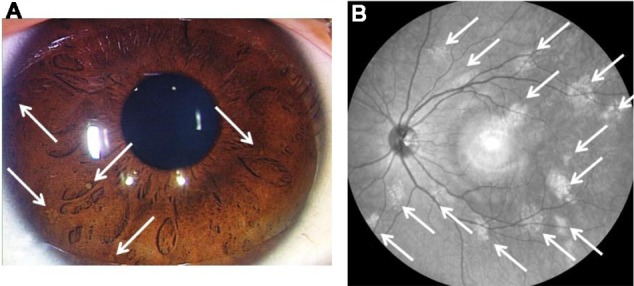
*[[File:Lisch nodules.jpg|alt=Lisch nodules and near-infrared reflectance image|thumb|Lisch nodules and near-infrared reflectance image (case 4). At least five Lisch nodules were detected and were classified as scale III (A). Note that 14 bright, patchy [[lesions]] were detected by near-infrared reflectance (B). The hyper-reflective point at the center of the image is an [[optical]] artifact. Case courtesy by Shinji Makino et al<ref>{{Cite web|url=https://www.ncbi.nlm.nih.gov/pmc/articles/PMC3883548/|title=Correlations between choroidal abnormalities, Lisch nodules, and age in patients with neurofibromatosis type 1|last=|first=|date=|website=|archive-url=|archive-date=|dead-url=|access-date=}}</ref>]]Lisch nodules occur in 90% of adults with [[Neurofibromatosis type I|neurofibromatosis]] 1.<ref name="pmid67892693">{{cite journal| author=Lewis RA, Riccardi VM| title=Von Recklinghausen neurofibromatosis. Incidence of iris hamartomata. | journal=Ophthalmology | year= 1981 | volume= 88 | issue= 4 | pages= 348-54 | pmid=6789269 | doi=10.1016/s0161-6420(81)35034-9 | pmc= | url=https://www.ncbi.nlm.nih.gov/entrez/eutils/elink.fcgi?dbfrom=pubmed&tool=sumsearch.org/cite&retmode=ref&cmd=prlinks&id=6789269 }}</ref><ref name="pmid31036733">{{cite journal| author=Huson S, Jones D, Beck L| title=Ophthalmic manifestations of neurofibromatosis. | journal=Br J Ophthalmol | year= 1987 | volume= 71 | issue= 3 | pages= 235-8 | pmid=3103673 | doi=10.1136/bjo.71.3.235 | pmc=1041127 | url=https://www.ncbi.nlm.nih.gov/entrez/eutils/elink.fcgi?dbfrom=pubmed&tool=sumsearch.org/cite&retmode=ref&cmd=prlinks&id=3103673 }}</ref><ref name="pmid289796203">{{cite journal| author=Abaloun Y, Ajhoun Y| title=[Lisch nodule in neurofibromatosis type 1]. | journal=Pan Afr Med J | year= 2017 | volume= 27 | issue= | pages= 218 | pmid=28979620 | doi=10.11604/pamj.2017.27.218.11517 | pmc=5622834 | url=https://www.ncbi.nlm.nih.gov/entrez/eutils/elink.fcgi?dbfrom=pubmed&tool=sumsearch.org/cite&retmode=ref&cmd=prlinks&id=28979620 }}</ref><ref name="pmid182803492">{{cite journal| author=Yang CC, Happle R, Chao SC, Yu-Yun Lee J, Chen W| title=Giant café-au-lait macule in neurofibromatosis 1: a type 2 segmental manifestation of neurofibromatosis 1? | journal=J Am Acad Dermatol | year= 2008 | volume= 58 | issue= 3 | pages= 493-7 | pmid=18280349 | doi=10.1016/j.jaad.2007.03.013 | pmc= | url=https://www.ncbi.nlm.nih.gov/entrez/eutils/elink.fcgi?dbfrom=pubmed&tool=sumsearch.org/cite&retmode=ref&cmd=prlinks&id=18280349 }}</ref><ref name="pmid204228424">{{cite journal| author=Cohen R, Shuper A| title=[Developmental manifestation in children with neurofibromatosis type 1]. | journal=Harefuah | year= 2010 | volume= 149 | issue= 1 | pages= 49-52, 61 | pmid=20422842 | doi= | pmc= | url=https://www.ncbi.nlm.nih.gov/entrez/eutils/elink.fcgi?dbfrom=pubmed&tool=sumsearch.org/cite&retmode=ref&cmd=prlinks&id=20422842 }}</ref><ref name="Dimitrova20094">{{cite journal|last1=Dimitrova|first1=Valentina|title=A CASE OF NEUROFIBROMATOSIS TYPE 1|journal=Journal of IMAB - Annual Proceeding (Scientific Papers)|volume=14, 1|issue=2008|year=2009|pages=63–67|issn=1312773X|doi=10.5272/jimab.14-1-2010.63}}</ref>
*On slit-lamp examination, they have a smooth, dome-shaped appearance and are usually light brown, although some can be very pale. <ref name="HusonKorf2013">{{cite journal|last1=Huson|first1=Susan M.|last2=Korf|first2=Bruce R.|title=The Phakomatoses|year=2013|pages=1–45|doi=10.1016/B978-0-12-383834-6.00128-2}}</ref>
*[[Eye]]-findings include orange-brown colored specks.
*Slit lamp examination can differentiate them from nevi on the iris by demonstrating elevated lesion instead of flat ones.
*Lisch nodules are usually elevated and tan in appearance.
*Lisch nodules develop during childhood, after the appearance of café-au-lait spots but before peripheral neurofibromas
*Lisch nodules are [[benign]] [[hamartomas]] that can be seen without magnification.
*This is useful in confirming the diagnosis of NF1 in children with no family history and only multiple café-au-lait spots
*Also known as [[Melanoma|melanocytic]] [[hamartomas]] of the iris, often associated with [[Neurofibromatosis type I|neurofibromatosis]] (NF) I.<ref name="pmid14560838">{{cite journal| author=Nichols JC, Amato JE, Chung SM| title=Characteristics of Lisch nodules in patients with neurofibromatosis type 1. | journal=J Pediatr Ophthalmol Strabismus | year= 2003 | volume= 40 | issue= 5 | pages= 293-6 | pmid=14560838 | doi= | pmc= | url=https://www.ncbi.nlm.nih.gov/entrez/eutils/elink.fcgi?dbfrom=pubmed&tool=sumsearch.org/cite&retmode=ref&cmd=prlinks&id=14560838 }}</ref>
*Other associated [[Ophthalmology|ophthalmologic]] findings are [[Optic glioma|optic gliomas]].
*[[Optic glioma|Optic gliomas]] can alter [[color vision]] and can produce progressive [[vision loss]].
==Diagnostic Studies==
*On [[slit-lamp]] examination, they have a smooth, dome-shaped appearance and are usually light brown, although some can be very pale.<ref name="pmid67892692">{{cite journal| author=Lewis RA, Riccardi VM| title=Von Recklinghausen neurofibromatosis. Incidence of iris hamartomata. | journal=Ophthalmology | year= 1981 | volume= 88 | issue= 4 | pages= 348-54 | pmid=6789269 | doi=10.1016/s0161-6420(81)35034-9 | pmc= | url=https://www.ncbi.nlm.nih.gov/entrez/eutils/elink.fcgi?dbfrom=pubmed&tool=sumsearch.org/cite&retmode=ref&cmd=prlinks&id=6789269 }}</ref><ref name="pmid31036734">{{cite journal| author=Huson S, Jones D, Beck L| title=Ophthalmic manifestations of neurofibromatosis. | journal=Br J Ophthalmol | year= 1987 | volume= 71 | issue= 3 | pages= 235-8 | pmid=3103673 | doi=10.1136/bjo.71.3.235 | pmc=1041127 | url=https://www.ncbi.nlm.nih.gov/entrez/eutils/elink.fcgi?dbfrom=pubmed&tool=sumsearch.org/cite&retmode=ref&cmd=prlinks&id=3103673 }}</ref><ref name="pmid289796204">{{cite journal| author=Abaloun Y, Ajhoun Y| title=[Lisch nodule in neurofibromatosis type 1]. | journal=Pan Afr Med J | year= 2017 | volume= 27 | issue= | pages= 218 | pmid=28979620 | doi=10.11604/pamj.2017.27.218.11517 | pmc=5622834 | url=https://www.ncbi.nlm.nih.gov/entrez/eutils/elink.fcgi?dbfrom=pubmed&tool=sumsearch.org/cite&retmode=ref&cmd=prlinks&id=28979620 }}</ref><ref name="pmid204228425">{{cite journal| author=Cohen R, Shuper A| title=[Developmental manifestation in children with neurofibromatosis type 1]. | journal=Harefuah | year= 2010 | volume= 149 | issue= 1 | pages= 49-52, 61 | pmid=20422842 | doi= | pmc= | url=https://www.ncbi.nlm.nih.gov/entrez/eutils/elink.fcgi?dbfrom=pubmed&tool=sumsearch.org/cite&retmode=ref&cmd=prlinks&id=20422842 }}</ref>
*[[Slit lamp]] examination can differentiate them from [[nevi]] on the [[iris]] by demonstrating elevated [[lesion]] instead of flat ones.<ref name="pmid19354164">{{cite journal| author=Crişan M, Talu S, Florea M, Coprean D, Cosgarea R, Crişan D| title=[Lisch nodules. Markers for a non-invasive diagnosis in Recklinghausen neurofibromatosis]. | journal=Oftalmologia | year= 2008 | volume= 52 | issue= 4 | pages= 56-61 | pmid=19354164 | doi= | pmc= | url=https://www.ncbi.nlm.nih.gov/entrez/eutils/elink.fcgi?dbfrom=pubmed&tool=sumsearch.org/cite&retmode=ref&cmd=prlinks&id=19354164 }}</ref>
*Lisch nodules develop during childhood, after the appearance of [[Café au lait spot|café-au-lait spots]] but before peripheral [[Neurofibroma|neurofibromas]].
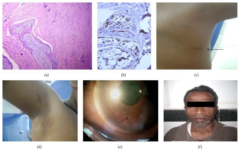
*[[File:Bundles of wavy spindle cells with serpentine nuclei .jpg|alt=Lisch nodule|thumb|(a) Microsection showing bundles of wavy [[spindle cells]] with serpentine nuclei in fascicles (H&E, ×10x), (b) Strong [[S-100 protein|S-100]] positivity of [[Tumor cell|tumor]] cells (×40x), (c) [[Café au lait spot|café au lait macule]] (arrow) in the back, (d) axillary [[freckle]], (e) Lisch nodule (arrow) in [[slit-lamp]] examination, and (f) father of patient with multiple cutaneous [[Neurofibroma|neurofibromas]]. Case courtesy by Rachna Rath et al.<ref>{{Cite web|url=https://www.ncbi.nlm.nih.gov/pmc/articles/PMC4921149/|title=Multifocal Head and Neck Neurofibromas with Osseous Abnormalities and Muscular Hypoplasia in a Child with Neurofibromatosis: Type I|last=|first=|date=|website=|archive-url=|archive-date=|dead-url=|access-date=}}</ref>]]This is useful in confirming the [[diagnosis]] of [[NF1]] in children with no [[family history]] and only multiple [[Café-au-lait spot|café-au-lait spots]].<ref name="pmid182803493">{{cite journal| author=Yang CC, Happle R, Chao SC, Yu-Yun Lee J, Chen W| title=Giant café-au-lait macule in neurofibromatosis 1: a type 2 segmental manifestation of neurofibromatosis 1? | journal=J Am Acad Dermatol | year= 2008 | volume= 58 | issue= 3 | pages= 493-7 | pmid=18280349 | doi=10.1016/j.jaad.2007.03.013 | pmc= | url=https://www.ncbi.nlm.nih.gov/entrez/eutils/elink.fcgi?dbfrom=pubmed&tool=sumsearch.org/cite&retmode=ref&cmd=prlinks&id=18280349 }}</ref><ref name="pmid196504184">{{cite journal| author=Terzi YK, Oguzkan-Balci S, Anlar B, Aysun S, Guran S, Ayter S| title=Reproductive decisions after prenatal diagnosis in neurofibromatosis type 1: importance of genetic counseling. | journal=Genet Couns | year= 2009 | volume= 20 | issue= 2 | pages= 195-202 | pmid=19650418 | doi= | pmc= | url=https://www.ncbi.nlm.nih.gov/entrez/eutils/elink.fcgi?dbfrom=pubmed&tool=sumsearch.org/cite&retmode=ref&cmd=prlinks&id=19650418 }}</ref>
==Treatment==
==Treatment==
{{CMG}}{{Swathi}}
=== Medical Therapy ===
*There is no treatment for the underlying disease nor any necessity to treat these small [[benign]] [[lesions]] which do not interfere with [[visual]] function.<ref name="Dimitrova20095">{{cite journal|last1=Dimitrova|first1=Valentina|title=A CASE OF NEUROFIBROMATOSIS TYPE 1|journal=Journal of IMAB - Annual Proceeding (Scientific Papers)|volume=14, 1|issue=2008|year=2009|pages=63–67|issn=1312773X|doi=10.5272/jimab.14-1-2010.63}}</ref>
*Lifelong monitoring is necessary because of the widespread manifestations and serious threat of [[complications]] such as:
**[[Visual]] [[impairment]]
**[[Renal]] [[hypertension]]
**[[Ischemia]] of major [[Organ (anatomy)|organs]].
== Primary Prevention ==
* There are no established measures for the [[primary prevention]] of Lisch nodules.
[[Lisch nodule medical therapy|Medical Therapy]]
== Secondary Prevention ==
*There is no treatment for the underlying disease nor any necessity to treat these small benign lesions which do not interfere with visual function, but lifelong monitoring is necessary because of the widespread manifestations and serious threat of complications such as visual impairment, renal hypertension and ischemia of major organs. <ref name="AdamsStewart2011">{{cite journal|last1=Adams|first1=E. G.|last2=Stewart|first2=K. M. A.|last3=Borges|first3=O. A.|last4=Darling|first4=T.|title=Multiple, Unilateral Lisch Nodules in the Absence of Other Manifestations of Neurofibromatosis Type 1|journal=Case Reports in Ophthalmological Medicine|volume=2011|year=2011|pages=1–2|issn=2090-6722|doi=10.1155/2011/854784}}</ref>
==Case Studies==
* There are no established measures for the [[secondary prevention]] of Lisch nodules.
In 1937, Karl Lisch published an article on the irishamartomas and their association with neurofibromatosis 1, now known as "Lisch nodules", while at the University Eye Clinic in Munich.
Lisch Nodules commonly associated with neurofibromatosis and is caused by genetic defects or mutations that either are passed on by a parent or occur spontaneously at conception.
The diagnosis is primarily based on clinical assessment and two or more of the features are required to confirm the diagnosis.
Physical Examination
Lisch nodules and near-infrared reflectance image (case 4). At least five Lisch nodules were detected and were classified as scale III (A). Note that 14 bright, patchy lesions were detected by near-infrared reflectance (B). The hyper-reflective point at the center of the image is an optical artifact. Case courtesy by Shinji Makino et al[38]Lisch nodules occur in 90% of adults with neurofibromatosis 1.[39][40][41][42][43][44]
↑Dimitrova, Valentina (2009). "A CASE OF NEUROFIBROMATOSIS TYPE 1". Journal of IMAB - Annual Proceeding (Scientific Papers). 14, 1 (2008): 63–67. doi:10.5272/jimab.14-1-2010.63. ISSN1312-773X.
↑Lubs, Marie-Louise E.; Bauer, Mislen S.; Formas, Maria E.; Djokic, Borivoje (1991). "Lisch Nodules in Neurofibromatosis Type 1". New England Journal of Medicine. 324 (18): 1264–1266. doi:10.1056/NEJM199105023241807. ISSN0028-4793.
↑Dimitrova, Valentina (2009). "A CASE OF NEUROFIBROMATOSIS TYPE 1". Journal of IMAB - Annual Proceeding (Scientific Papers). 14, 1 (2008): 63–67. doi:10.5272/jimab.14-1-2010.63. ISSN1312-773X.
↑Richetta, A; Giustini, S; Recupero, SM; Pezza, M; Carlomagno, V; Amoruso, G; Calvieri, S (2004). "Lisch nodules of the iris in neurofibromatosis type 1". Journal of the European Academy of Dermatology and Venereology. 18 (3): 342–344. doi:10.1111/j.1468-3083.2004.00915.x. ISSN0926-9959.
↑Lubs, Marie-Louise E.; Bauer, Mislen S.; Formas, Maria E.; Djokic, Borivoje (1991). "Lisch Nodules in Neurofibromatosis Type 1". New England Journal of Medicine. 324 (18): 1264–1266. doi:10.1056/NEJM199105023241807. ISSN0028-4793.
↑Lubs, Marie-Louise E.; Bauer, Mislen S.; Formas, Maria E.; Djokic, Borivoje (1991). "Lisch Nodules in Neurofibromatosis Type 1". New England Journal of Medicine. 324 (18): 1264–1266. doi:10.1056/NEJM199105023241807. ISSN0028-4793.
↑Dimitrova, Valentina (2009). "A CASE OF NEUROFIBROMATOSIS TYPE 1". Journal of IMAB - Annual Proceeding (Scientific Papers). 14, 1 (2008): 63–67. doi:10.5272/jimab.14-1-2010.63. ISSN1312-773X.
↑Dimitrova, Valentina (2009). "A CASE OF NEUROFIBROMATOSIS TYPE 1". Journal of IMAB - Annual Proceeding (Scientific Papers). 14, 1 (2008): 63–67. doi:10.5272/jimab.14-1-2010.63. ISSN1312-773X.
↑Dimitrova, Valentina (2009). "A CASE OF NEUROFIBROMATOSIS TYPE 1". Journal of IMAB - Annual Proceeding (Scientific Papers). 14, 1 (2008): 63–67. doi:10.5272/jimab.14-1-2010.63. ISSN1312-773X.