Acute lymphoblastic leukemia classification: Difference between revisions
Carlos Lopez (talk | contribs) |
Carlos Lopez (talk | contribs) |
||
| Line 40: | Line 40: | ||
{| align="center" | {| align="center" | ||
|-valign="top" | |-valign="top" | ||
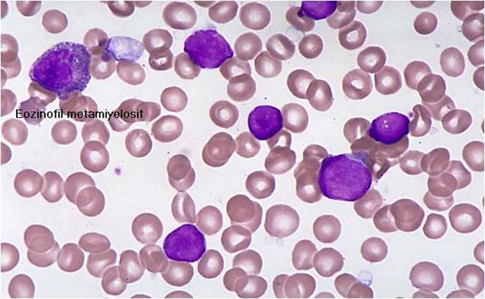
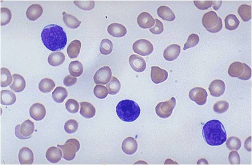
| [[Image:ALL-L1.jpg|thumb| | | [[Image:ALL-L1.jpg|thumb|Acute lymphoblastic leukemia -L1: Small uniform cells subtype]] | ||
| [[Image:ALL-L1 0002.jpg|thumb| | | [[Image:ALL-L1 0002.jpg|thumb|Acute lymphoblastic leukemia-L1: Large varied cells subtype]] | ||
|} | |} | ||
| Line 47: | Line 47: | ||
{| align="center" | {| align="center" | ||
|-valign="top" | |-valign="top" | ||
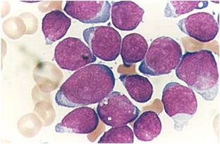
| [[Image:ALL-L2 Hand mirror cell 0001.jpg|thumb| | | [[Image:ALL-L2 Hand mirror cell 0001.jpg|thumb|Acute lymphoblastic leukemia-L2: Large varied cells (Hand mirror cell)]] | ||
| [[Image:ALL-L2 0003.jpg|thumb| | | [[Image:ALL-L2 0003.jpg|thumb|Acute lymphoblastic leukemia-L2: Large varied cells]] | ||
|} | |} | ||
| Line 54: | Line 54: | ||
{| align="center" | {| align="center" | ||
|-valign="top" | |-valign="top" | ||
| [[Image:ALL-L2 0002.jpg|thumb| | | [[Image:ALL-L2 0002.jpg|thumb|Acute lymphoblastic leukemia-L2: Large varied cells]] | ||
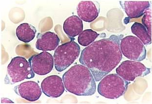
| [[Image:ALL-L3 0001.jpg|thumb| | | [[Image:ALL-L3 0001.jpg|thumb|Acute lymphoblastic leukemia-L3: Large varied cells with vacuoles (bubble-like features]] | ||
|} | |} | ||
Revision as of 19:50, 28 August 2015
|
Acute lymphoblastic leukemia Microchapters |
|
Differentiating Acute lymphoblastic leukemia from other Diseases |
|---|
|
Diagnosis |
|
Treatment |
|
Case Studies |
|
Acute lymphoblastic leukemia classification On the Web |
|
American Roentgen Ray Society Images of Acute lymphoblastic leukemia classification |
|
Directions to Hospitals Treating Acute lymphoblastic leukemia |
|
Risk calculators and risk factors for Acute lymphoblastic leukemia classification |
Editor-In-Chief: C. Michael Gibson, M.S., M.D. [1] Associate Editor(s)-in-Chief: Raviteja Guddeti, M.B.B.S. [2] Carlos A Lopez, M.D. [3]
Overview
Acute lymphoblastic leukemia may be classified according to the French-American-British classification (FAB) and World Health Organization classification (WHO). The French-American-British classification is divided into 3 groups: ALL-L1: small uniform cells, ALL-L2: large varied cells, ALL-L3: large varied cells with vacuoles (bubble-like features). The World Health Organization classification is divided into three 3 too: B lymphoblastic leukemia/lymphoma, B lymphoblastic leukemia/lymphoma (Not organ specific), B lymphoblastic leukemia/lymphoma with recurrent genetic abnormalities.
Classification
There are 2 classifications systems for acute lymphoblastic leukemia about acute lymphoblastic leukemia, the French-American-British classification and the "World Health Organization":
French-American-British classification
The French-American-British (FAB) classification of acute lymphoblastic leukemia is divided into 3 subtypes L1 to L3 based on the type of cell from which the leukemia developed and its degree of maturity and morphological classification. Each subtype is then further classified by determining the surface markers of the abnormal lymphocytes, called immunophenotyping. There are 2 main immunologic types: pre-B cell and pre-T cell. The mature B-cell acute lymphoblastic leukemia (L3) is now classified as Burkitt leukemia/lymphoma.
Classification
- ALL-L1: small uniform cells
- ALL-L2: large varied cells
- ALL-L3: large varied cells with vacuoles (bubble-like features)
World Health Organization classification
The World Health Organization (WHO) classification of acute lymphoblastic leukemia attempts to be more clinically useful and to produce more meaningful prognostic information than the French-American-British (FAB) classification criteria. There are 3 different grups of Lymphoblastic leukemias according to the WHO including a classification with recurrent genetic abnormalities.[1]
Group 1
- B lymphoblastic leukemia/lymphoma
Group 2
- B lymphoblastic leukemia/lymphoma (Not organ specific)
Group 3
- B lymphoblastic leukemia/lymphoma with recurrent genetic abnormalities:
- B lymphoblastic leukemia/lymphoma with t(9;22)(q34;q11.2), BCR-ABL1
- B lymphoblastic leukemia/lymphoma with t(v;11q23); MLL rearranged
- B lymphoblastic leukemia/lymphoma with t(12;21)(p13;q22) TEL-AML1 (ETV6-RUNX1)
- B lymphoblastic leukemia/lymphoma with hyperdiploidy
- B lymphoblastic leukemia/lymphoma with hypodiploidy
- B lymphoblastic leukemia/lymphoma with t(5;14)(q31;q32) IL3-IGH
- B lymphoblastic leukemia/lymphoma with t(1;19)(q23;p13.3) TCF3-PBX1
(Images shown below are courtesy of Melih Aktan MD, Istanbul Medical Faculty - Turkey, and Kyoto University - Japan)
 |
 |
 |
 |
 |
 |
References
- ↑ Campo E, Swerdlow SH, Harris NL, Pileri S, Stein H, Jaffe ES (2011). "The 2008 WHO classification of lymphoid neoplasms and beyond: evolving concepts and practical applications". Blood. 117 (19): 5019–32. doi:10.1182/blood-2011-01-293050. PMC 3109529. PMID 21300984.