Abdominal pain differential diagnosis

Jump to navigation Jump to search
<figure-inline><figure-inline><figure-inline><figure-inline><figure-inline></figure-inline></figure-inline></figure-inline></figure-inline></figure-inline>

Resident
Survival
Guide

Abdominal pain Microchapters

Home

Patient Information

Overview

Classification

Pathophysiology

Causes

Diffuse Abdominal Pain
Left Upper Quadrant
Left Flank
Left Lower Quadrant
Epigastric Quadrant
Umbilical Region
Pelvic Region
Right Upper Quadrant
Right Flank
Right Lower Quadrant
Intraperitoneal
Extraperitoneal

Differentiating Abdominal pain from other Diseases

Epidemiology and Demographics

Risk Factors

Screening

Natural History, Complications and Prognosis

Diagnosis

History and Symptoms

Physical Examination

Laboratory Findings

Electrocardiogram

X Ray

CT

Other Imaging Findings

Other Diagnostic Studies

Treatment

Medical Therapy

Surgery

Primary Prevention

Secondary Prevention

Cost-Effectiveness of Therapy

Future or Investigational Therapies

Case Studies

Case #1

Abdominal pain On the Web

Most recent articles

Most cited articles

Review articles

CME Programs

Powerpoint slides

Google Images

American Roentgen Ray Society Images of Abdominal pain

All Images
X-rays
Echo & Ultrasound
CT Images
MRI

Ongoing Trials at Clinical Trials.gov

US National Guidelines Clearinghouse

NICE Guidance

FDA on Abdominal pain

CDC on Abdominal pain

Abdominal pain in the news

Blogs on Abdominal pain

Directions to Hospitals Treating Abdominal pain

Risk calculators and risk factors for Abdominal pain

Editor-In-Chief: C. Michael Gibson, M.S., M.D. [1];Associate Editor(s)-in-Chief: Seyedmahdi Pahlavani, M.D. [2]Amandeep Singh M.D.[3]

Overview

Diagnosing the cause of abdominal pain can be difficult, because many diseases can cause this symptom. Most frequently the cause is benign and/or self-limiting, but more serious causes may require urgent intervention. Acute abdominal pain is a severe, persistent abdominal pain of sudden onset that is likely to require surgical intervention to treat its cause. The following table summarizes differential diagnosis for abdominal pain.

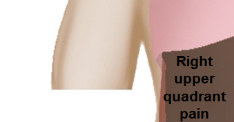
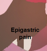
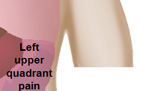
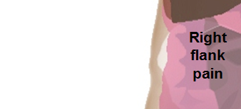
Differential diagnosis of abdominal pain

Classification of acute abdomen based on etiology Disease Clinical manifestations Diagnosis Comments
Symptoms Signs
Fever Rigors and chills Abdominal Pain Jaundice Hypo-

tension

Guarding Rebound Tenderness Bowel sounds Lab Findings Imaging
Abdominal causes Inflammatory causes Pancreato-biliary disorders Acute suppurative cholangitis + + RUQ + + + + N
  • Abnormal LFT
  • WBC >10,000
Ultrasound shows biliary dilatation/stents/tumor Septic shock occurs with features of SIRS
Acute cholangitis + RUQ + N Abnormal LFT Ultrasound shows biliary dilatation/stents/tumor Biliary drainage (ERCP) + IV antibiotics
Acute cholecystitis + RUQ + Hypoactive Ultrasound shows gallstone and evidence of inflammation Murphy’s sign
Acute pancreatitis + Epigastric ± ± N Increased amylase / lipase Ultrasound shows evidence of inflammation Pain radiation to back
Primary biliary cirrhosis RUQ/Epigastric + N Increased AMA level, abnormal LFTs
Primary sclerosing cholangitis + RUQ + N
Cholelithiasis ± RUQ/Epigastric ± + + N to hyperactive for dislodged stone Leukocytosis Ultrasound shows gallstone Murphy’s sign
Gastric causes Peptic ulcer disease ± Diffuse + in perforated + + N
  • Ascitic fluid
    • LDH > serum LDH
    • Glucose < 50mg/dl
    • Total protein > 1g/dl
Air under diaphragm in upright CXR Upper GI endoscopy for diagnosis
Gastritis ± Epigastric
Gastroesophageal reflux disease Epigastric
Gastric outlet obstruction Epigastric ± Hyperactive
Gastrointestinal perforation + ± Diffuse ± + + ± WBC> 10,000 Air under diaphragm in upright CXR
Intestinal causes Acute appendicitis + +in pyogenic appendicitis Starts in epigastrium, migrates to RLQ + in perforated appendicitis + + Hypoactive Leukocytosis Ultrasound shows evidence of inflammation Nausea & vomiting, decreased appetite
Acute diverticulitis + ± LLQ + Hypoactive Leukocytosis CT scan and ultrasound shows evidence of inflammation
Inflammatory bowel disease ± Diffuse
Irritable bowel syndrome ± Diffuse N Tests done to exclude other diseases as it diagnosis of exclusion Tests done to exclude other diseases as it diagnosis of exclusion Symptomatic treatment
Whipple's disease ± Diffuse ± ± N *Endoscopy is used to confirm diagnosis.

Images used to find complications

Extra intestinal findings:
Toxic megacolon + Diffuse + ± Hypoactive
Tropical sprue + Diffuse
Celiac disease Diffuse ±, also dermatitis herpetiformis Hyperactive (increased sounds)
Hepatic causes Viral hepatitis + RUQ + +
Liver masses + + in Liver abscess RUQ ± + in sepsis
Budd-Chiari syndrome ± RUQ
Hemochromatosis RUQ Dull / aching + in cirrhotic patients may be in cardicmyopathy >60% TS
>240 μg/L SF
Raised LFT
Hyperglycemia
Ultrasound shows evidence of cirrhosis Extra intestinal findings:
  • hyperpigmentation
  • Diabetes mellitus
  • Arthralgia
  • Impotence in males
  • Cardiomyopathy
  • Atherosclerosis
  • Hypopituitarism
  • Hypothyroidism
  • Extrahepatic cancer
  • Prone to specific infections
Cirrhosis + RUQ +
Peritoneal causes Spontaneous bacterial peritonitis + Diffuse + in cirrhotic patients ± Hypoactive
  • Ascitic fluid PMN>250 cells/mm³
  • Culture: Positive for single organism
Ultrasound for evaluation of liver cirrhosis
Hollow Viscous Obstruction Small intestine obstruction Diffuse + ± Hyperactive then absent Leukocytosis Abdominal X ray Nausea & vomiting associated with constipation, abdominal distention
Volvulus Diffuse + Hypoactive Leukocytosis CT scan and abdominal X ray Nausea & vomiting associated with constipation, abdominal distention
Biliary colic RUQ + N Increased bilirubin and alkaline phosphatase Ultrasound Nausea & vomiting
Renal colic Flank pain N Hematuria CT scan and ultrasound Colicky abdominal pain associated with nausea & vomiting
Vascular Disorders Ischemic causes Mesenteric ischemia ± Periumbilical ± Hyperactive Leukocytosis and lactic acidosis CT scan Nausea & vomiting, normal physical examination
Acute ischemic colitis ± ± Diffuse + + Hyperactive then absent Leukocytosis CT scan Nausea & vomiting
Hemorrhagic causes Ruptured abdominal aortic aneurysm Diffuse + N Normal CT scan Unstable hemodynamics
Intra-abdominal or retroperitoneal hemorrhage Diffuse + N Anemia CT scan History of trauma
Gynaecological Causes Tubal causes Torsion of the cyst RLQ / LLQ ± ± N Increased ESR and CRP Ultrasound Sudden onset sever pain with nausea and vomiting
Acute salpingitis + ± RLQ / LLQ ± ± N Leukocytosis Pelvic ultrasound Vaginal discharge
Cyst rupture RLQ / LLQ + ± ± N Increased ESR and CRP Ultrasound Sudden onset sever pain with nausea and vomiting
Pregnancy Ruptured ectopic pregnancy RLQ / LLQ + N Positive pregnancy test Ultrasound History of missed period and vaginal bleeding
Extra-abdominal causes Pulmonary disorders Pleural empyema + ± RUQ/Epigastric N
Cardiovascular disorders Myocardial Infarction Epigastric + in cardiogenic shock N


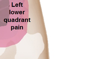
The following is a list of diseases that present with acute onset severe lower abdominal pain:

Disease Findings
Ectopic pregnancy History of missed menses, positive pregnancy test, ultrasound reveals an empty uterus and may show a mass in the fallopian tubes.[1]
Appendicitis Pain localized to the right iliac fossa, vomiting, abdominal ultrasound sensitivity for diagnosis of acute appendicitis is 75% to 90%.[2]
Rupturedovarian cyst Usually spontaneous, can follow history of trauma, mild chronic lower abdominal discomfort may suddenly intensify, ultrasound is diagnostic.[3]
Ovarian cyst torsion Presents with acute severe unilateral lower quadrant abdominal pain, nausea and vomiting, tender adnexal mass palpated in 90%, ultrasound is diagnostic.[4]
Hemorrhagic ovarian cyst Presents with localized abdominal pain, nausea and vomiting. Hypovolemic shock may be present, abdominal tenderness and guarding are physical exam findings, ultrasound is diagnostic.[4]
Endometriosis Presents with cyclic pain that is exacerbated by onset of menses, dyspareunia. laparoscopic exploration is diagnostic.[4]
Acute cystitis Presents with features of increased urinary frequency, urgency, dysuria, and suprapubic pain.[5][6]

References

  1. Morin L, Cargill YM, Glanc P (2016). "Ultrasound Evaluation of First Trimester Complications of Pregnancy". J Obstet Gynaecol Can. 38 (10): 982–988. doi:10.1016/j.jogc.2016.06.001. PMID 27720100.
  2. Balthazar EJ, Birnbaum BA, Yee J, Megibow AJ, Roshkow J, Gray C (1994). "Acute appendicitis: CT and US correlation in 100 patients". Radiology. 190 (1): 31–5. doi:10.1148/radiology.190.1.8259423. PMID 8259423.
  3. Bottomley C, Bourne T (2009). "Diagnosis and management of ovarian cyst accidents". Best Pract Res Clin Obstet Gynaecol. 23 (5): 711–24. doi:10.1016/j.bpobgyn.2009.02.001. PMID 19299205.
  4. 4.0 4.1 4.2 Bhavsar AK, Gelner EJ, Shorma T (2016). "Common Questions About the Evaluation of Acute Pelvic Pain". Am Fam Physician. 93 (1): 41–8. PMID 26760839.
  5. {{Cite journal | author = W. E. Stamm | title = Etiology and management of the acute urethral syndrome | journal = Sexually transmitted diseases | volume = 8 | issue = 3 | pages = 235–238 | year = 1981 | month = July-September | pmid = 7292216
  6. {{Cite journal | author = W. E. Stamm, K. F. Wagner, R. Amsel, E. R. Alexander, M. Turck, G. W. Counts & K. K. Holmes | title = Causes of the acute urethral syndrome in women | journal = The New England journal of medicine | volume = 303 | issue = 8 | pages = 409–415 | year = 1980 | month = August | doi = 10.1056/NEJM198008213030801 | pmid = 6993946

Template:Gastroenterology


Template:WikiDoc Sources |}