Endocarditis electrocardiogram
|
Endocarditis Microchapters |
|
Diagnosis |
|---|
|
Treatment |
|
2014 AHA/ACC Guideline for the Management of Patients With Valvular Heart Disease |
|
Case Studies |
|
Endocarditis electrocardiogram On the Web |
|
Risk calculators and risk factors for Endocarditis electrocardiogram |
Editor-In-Chief: C. Michael Gibson, M.S., M.D. [1]; Associate Editors-in-Chief: Cafer Zorkun, M.D., Ph.D. [2]
Overview
There is no specific EKG changes that are diagnostic of Infective Endocarditis.
Electrocardiogram
The EKG may be useful in the detection of the 10% of patients who develop a conduction delay during Infective Endocarditis by documenting an increased PR interval. If myocardial infarction is present, it may be due to vessel occlusion with ST elevation myocardial infarction or it may be due to distal embolism which may result in non ST elevation MI.
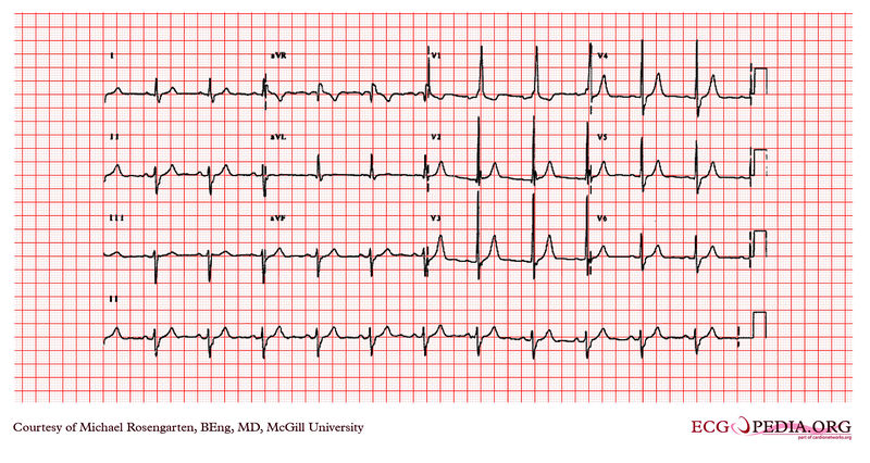
This is an electrocardiogram from a man in his 80's. The patient has severe lung disease, has mitral regurgitation secondary to bacterial endocarditis , and is taking digoxin, Lasix and potassium.
The electrocardiogram shows sinus rhythm and a QRS with a left axis deviation, a QRS duration of 118 milliseconds and a tall R wave in the first precordial lead V1 with an R wave height of approximately 21 mm. The prolonged QRS duration and the S waves that are seen as lead 1 and lead 6 suggest a right on the branch block and the a left axis deviation suggests a left anterior hemi-block . Finally the tall R wave in V1 lead suggests right ventricular hypertrophy.
