Turner syndrome physical examination
Jump to navigation
Jump to search
|
Turner syndrome Microchapters |
|
Diagnosis |
|---|
|
Treatment |
|
Case Studies |
|
Turner syndrome physical examination On the Web |
|
American Roentgen Ray Society Images of Turner syndrome physical examination |
|
Risk calculators and risk factors for Turner syndrome physical examination |
Editor-In-Chief: C. Michael Gibson, M.S., M.D. [1]; Associate Editor(s)-in-Chief: Akash Daswaney, M.B.B.S[2]
Overview
Physical examination may be suggestive of thyroid dysfunction, congenital heart defects, inflammatory bowel disease, characteristic skeletal deformities and body habitus/skin manifestations.
Physical Examination
Appearance of the Patient
- Patient may show signs of poor growth velocity and malnutrition indicative of failure to thrive.
- Patients may be hyperactive.
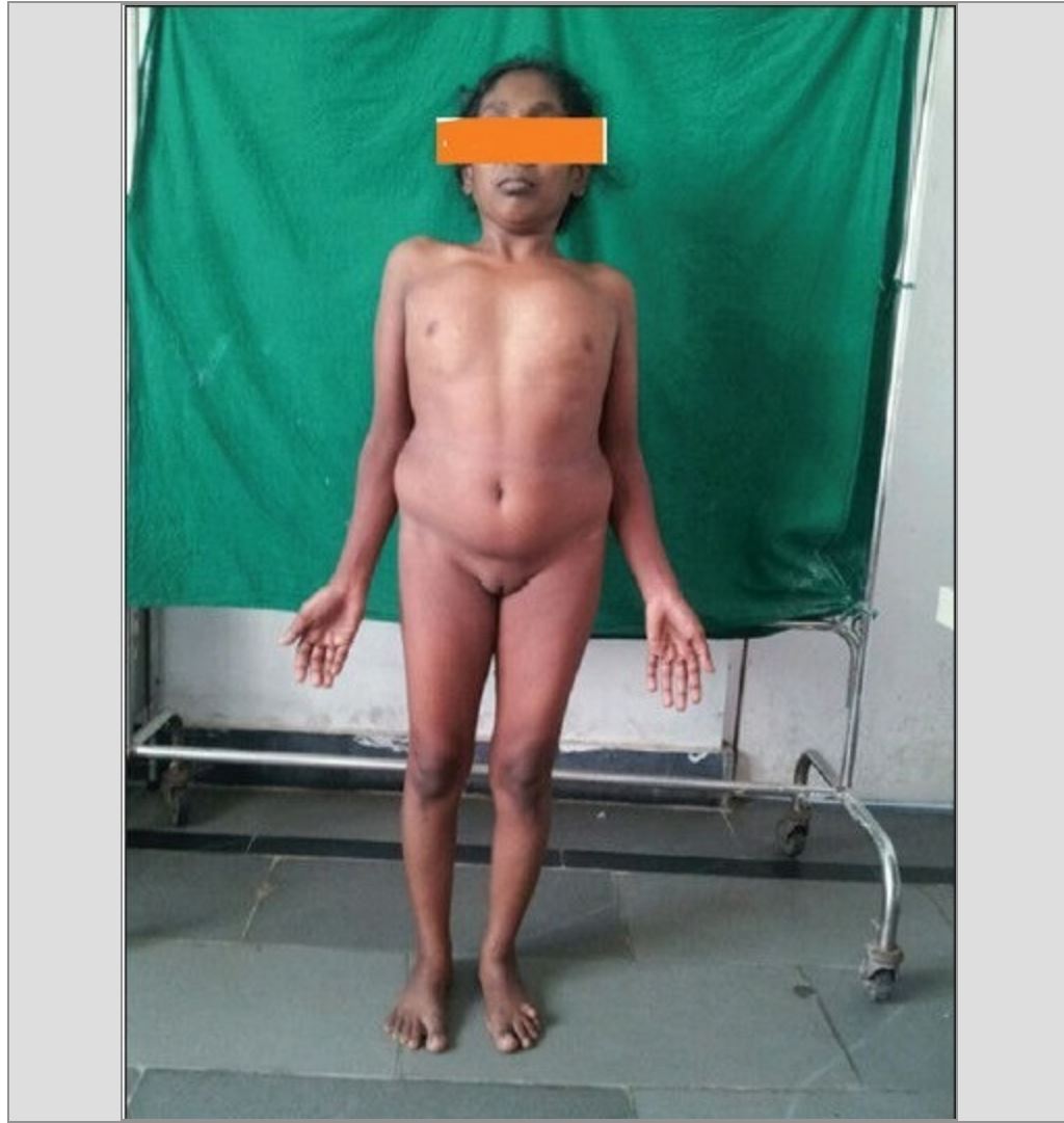
- The presence of short stature, a webbed neck, pigmented nevi, puffiness of the hands and feet, a cubitus valgus deformity, genu valgum on physical examination are highly suggestive of Turner syndrome.
Vital Signs
- High-grade / low-grade fever
- Hypothermia / hyperthermia may be present
- Tachycardia with regular pulse or irregularly irregular pulse - Hyperthyroidism
- Bradycardia with regular pulse or irregularly irregular pulse - Hypothyroidism
- Tachypnea - Secondary to Acute pulmonary edema from ischemic heart disease
- Hypertension in the upper extremities with hypotension in the lower extremities, decreased post ductal oxygen saturation in the lower extremities, absent lower extremity pulses - Coarctation of aorta
- Widened pulse pressure + water hammer pulse - Aortic Regurgitation secondary to aortic dissection
Skin
- Lymphedema of hands and feet
- Toe nail cellulitis
- Vitiligo
- Alopecia
- Nail hypoplasia
- Psoriasis - silver scaled erythrematous plaques present on extensor surfaces
- Pigmented melanocytic nevi - avoid rubbing against clothes.
- Hyperconvex nails
- Oslers nodes, Jane way lesions]] - Infective Endocarditis
HEENT
Ear
- Weber test may be abnormal- Conductive hearing loss or sensorineural hearing loss
- Rinne test may be positive - Conductive hearing loss or sensorineural hearing loss
- External ear canal deformities may be noted.
Eye
- Prominent epicanthal folds
- Bilateral epicanthus
- Strabismus
- Ptosis
- Cataract
- Nystagmus
- Roth spots - secondary to infective endocarditis
Neck
- Pterygium colli
- Low posterior hair line
- Loose skin on the nape of newborns
- Jugular venous distension - Rule out Eisenmengirsation secondary to congenital heart defects, pressure may be transmitted to the internal jugular vein.
- Hepatojugular reflux - There is an increased risk of ischemic heart disease and cardiomyopathy secondary to this may cause Right sided heart failure.
Lungs
- Pulmonary examination of patients with Turner syndrome is usually normal.
- Fine/coarse crackles upon auscultation of the lung bases/apices unilaterally/bilaterally (rule out)
Heart
- Patient has a wide shield shaped chest with inverted nipples.
- S3 (rule out)
- S4 (rule out)
- Gallops (rule out)
- A pansystolic murmur heard over the tricuspid area - Ventricular Septal defect
- Early blowing diastolic murmur head over the left upper sternal border - Aortic regurgitation secondary to aortic dissection
- Systolic ejection click followed by a crescendo decrescendo murmur (early P2 and delayed A2) - Early onset aortic stenosis secondary to a bicuspid aortic valve
- Fever + Petechiae + Heart murmur - Infective Endocarditis
Abdomen
- Look for signs of inflammatory bowel disease such as fistulas, skin tags and oral aphtous ulcers
Back
Genitourinary
- Chronic estrogen deficiency may lead to signs of atrophic vaginitis
- Rudimentary uterus
- Palpable mass secondary to dysgerminoma or gonadoblastoma
Neuromuscular
- Neuromuscular examination of patients with Turner syndrome is usually normal.
Extremities
- Shortened limbs
- Shortened 4th metacarpal
- Cubitus valgus deformity
- Madelung deformity
- Cyanosis - Secondary to structural heart defects
- Pitting edema of the upper/lower extremities - Hypothyroidism
- Muscle atrophy
References
- ↑ Bucerzan S, Miclea D, Popp R, Alkhzouz C, Lazea C, Pop IV; et al. (2017). "Clinical and genetic characteristics in a group of 45 patients with Turner syndrome (monocentric study)". Ther Clin Risk Manag. 13: 613–622. doi:10.2147/TCRM.S126301. PMC 5422538. PMID 28496331.
- ↑ 2.0 2.1 Vaddadi S, Murthy RS, Rahul CH, Kumar VL (2013). "A rare case of Turner's syndrome presenting with Mullerian agenesis". J Hum Reprod Sci. 6 (4): 277–9. doi:10.4103/0974-1208.126313. PMC 3963314. PMID 24672170.
![[1]](/images/c/c7/Turner_Syndrome_Phenotype.JPG)
![[2]](/images/0/03/Turner_Syndrome_Phenotype_2.JPG)
![[2]](/images/7/77/Turner_Syndrome_Phenotype_3.JPG)
