Subarachnoid hemorrhage CT
|
Subarachnoid Hemorrhage Microchapters |
|
Diagnosis |
|---|
|
Treatment |
|
AHA/ASA Guidelines for the Management of Aneurysmal Subarachnoid Hemorrhage (2012)
|
|
Case Studies |
|
Subarachnoid hemorrhage CT On the Web |
|
American Roentgen Ray Society Images of Subarachnoid hemorrhage CT |
|
Risk calculators and risk factors for Subarachnoid hemorrhage CT |
Editor-In-Chief: C. Michael Gibson, M.S., M.D. [1]; Associate Editor(s)-In-Chief: Cafer Zorkun, M.D., Ph.D. [2]
CT
- Subarachnoid hemorrhage appears as a high-attenuating, amorphous substance that fills the normally dark CSF-filled subarachnoid spaces.
- These findings are most evident in the largest subarachnoid spaces, such as the suprasellar cistern and Sylvian fissures.
- Acute Subarachnoid hemorrhage is typically 50-60 HU.
- When CT scanning is performed several days to weeks after the initial bleed, the findings are more subtle.
- The initial high-attenuation of blood and clot tend to decrease, and these appear as intermediate gray.
- These findings can be isointense relative to normal brain parenchyma.
- In addition to detecting Subarachnoid hemorrhage, CT is useful in localizing the source of bleeding.
-
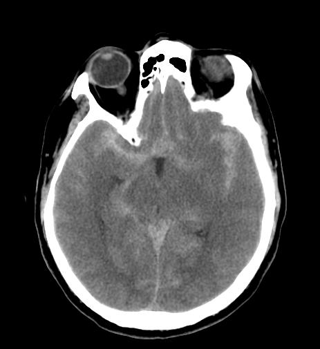
CT: Diffuse subarachnoid hemorrhage
-
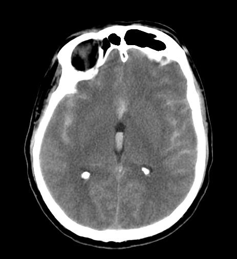
CT: Diffuse subarachnoid hemorrhage
-
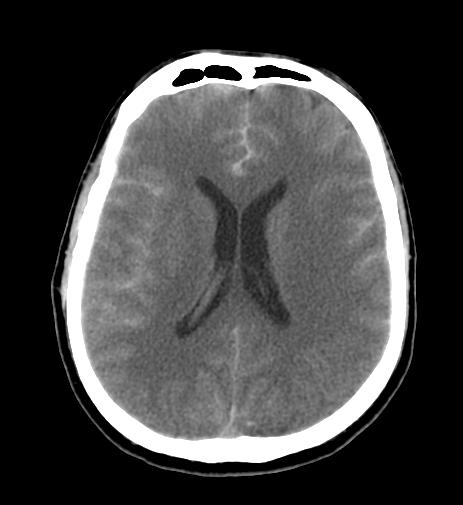
CT: Diffuse subarachnoid hemorrhage
-
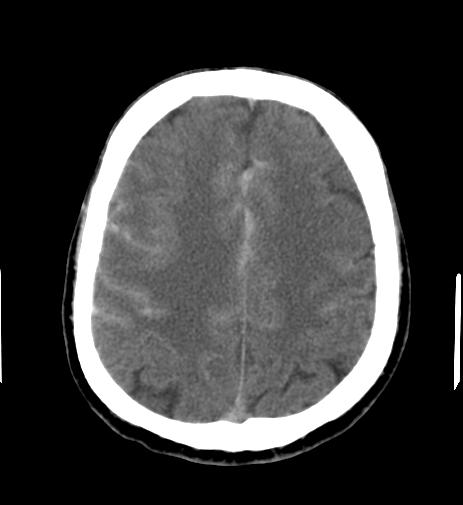
CT: Diffuse subarachnoid hemorrhage


