Angiomyolipoma: Difference between revisions
No edit summary |
No edit summary |
||
| Line 119: | Line 119: | ||
[[Category:Oncology]] | [[Category:Oncology]] | ||
[[Category:Disease]] | |||
[[de:Angiomyolipom]] | [[de:Angiomyolipom]] | ||
[[fr:Angiomyolipome]] | [[fr:Angiomyolipome]] | ||
Revision as of 13:16, 11 September 2012
| Angiomyolipoma | |
 | |
|---|---|
| ICD-10 | D30.0 |
| ICD-9 | 223.0 |
| ICD-O: | M8860/0 |
| DiseasesDB | 29496 |
| MeSH | D018207 |
|
Angiomyolipoma Microchapters |
|
Diagnosis |
|---|
|
Treatment |
|
Case Studies |
|
Angiomyolipoma On the Web |
|
American Roentgen Ray Society Images of Angiomyolipoma |
Editor-In-Chief: C. Michael Gibson, M.S., M.D. [1] Associate Editor-In-Chief: Cafer Zorkun, M.D., Ph.D. [2]
Overview
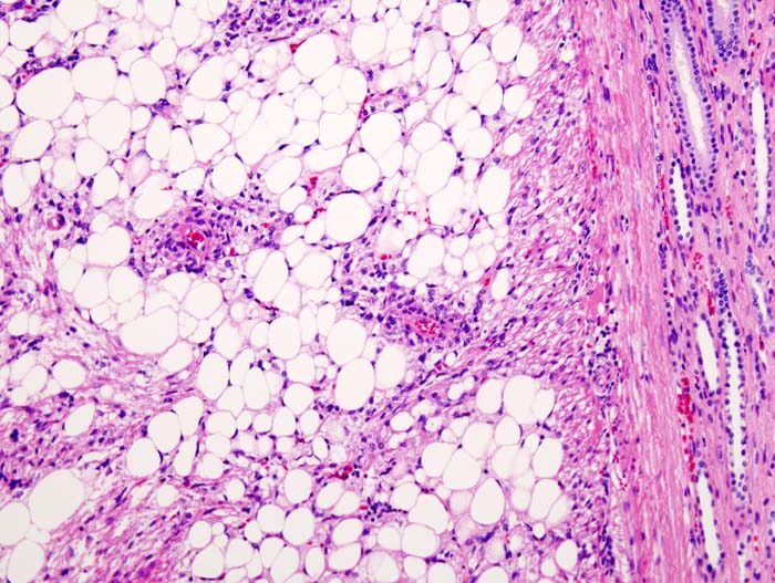
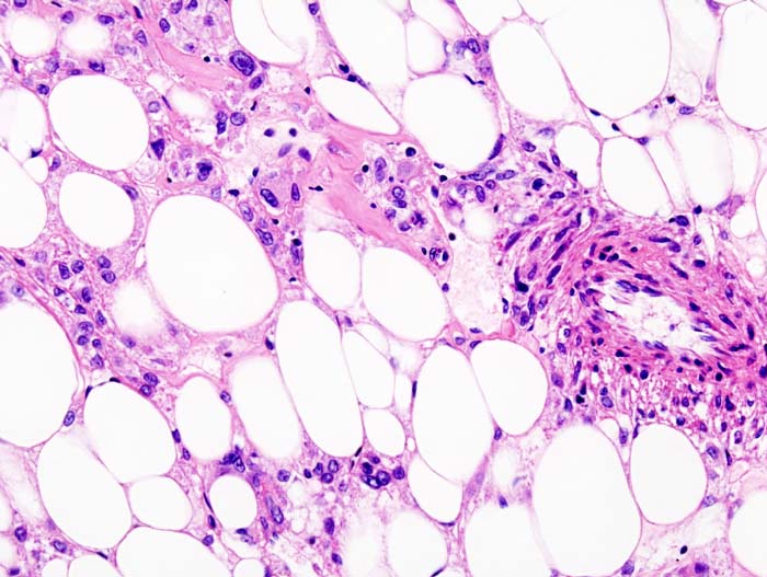
Angiomyolipoma is a benign renal neoplasm previously considered to be a hamartoma or choristoma, but now known to be neoplastic.[1] It is composed of variable amounts of fat, vascular, and smooth muscle elements. The fat density of the tumour on CT has been regarded to be pathognomonic, although there are now case reports of renal cell carcinoma types also possessing macroscopic fat.
The lesion is well demarcated and contains mature elements. It occurs in more than 50% of individuals with tuberous sclerosis, often bilaterally. Angiomyolipomata also occur in 40% of women who have a rare, cystic lung disease called lymphangioleiomyomatosis, or LAM.[2]
Incidence
The incidence is about 0.3-3%. It occurs in more than 50% of individuals with tuberous sclerosis, often bilaterally. It is twice as common in females as in males.
Types
- Isolated Angiomyolipoma (80%)
- Angiomyolipoma is about 4 times more common in women than in men.
- Interestingly, 80% of the cases involve the right kidney.
- Angiomyolipoma associated with tuberous sclerosis (20%).
- Angiomyolipoma occurs in 80% patients with tuberous sclerosis.
- Angiomyolipoma also occurs young women with lymphangiomyomatosis without other stigmata of tuberous sclerosis. Angiomyolipoma and lymphangiomyomatosis are sometimes considered the forme fruste of tuberous sclerosis.
Diagnosis
Demonstration of fat in renal tumor is virtually diagnostic of angiomyolipoma. Most small lesions are asymptomatic and incidental findings on images. The fat density of the tumour on CT is pathognomonic.
Diagnostic Findings
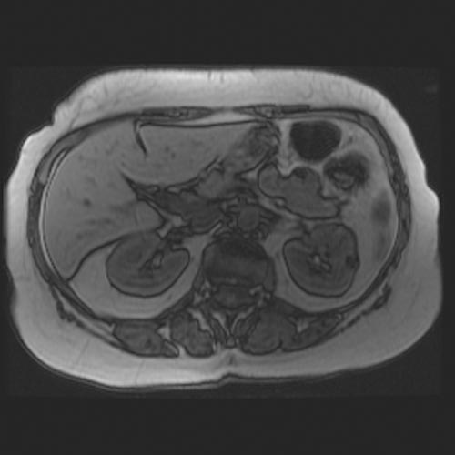
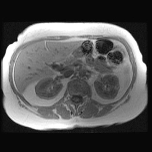
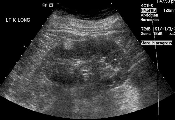
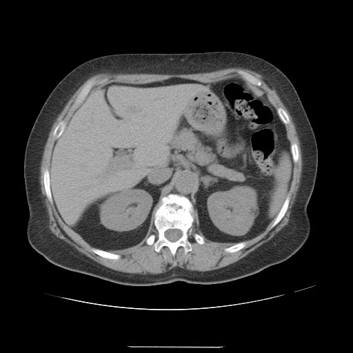
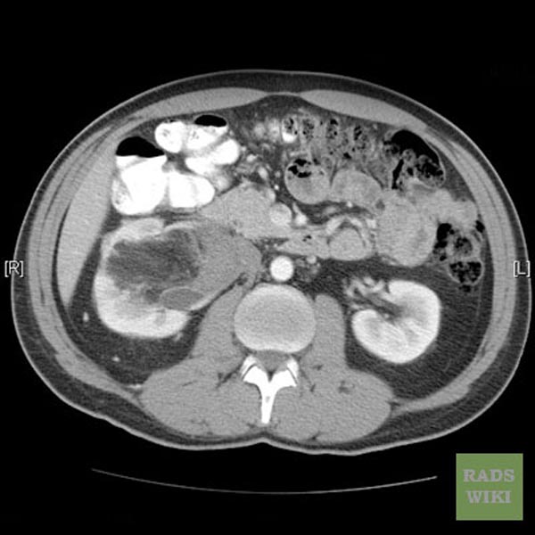
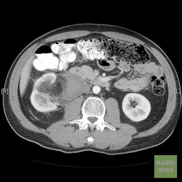
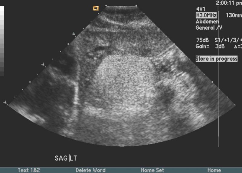
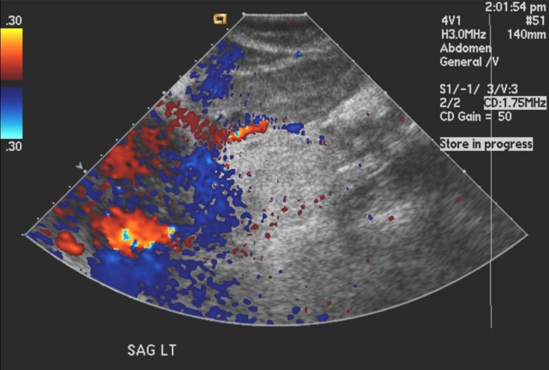
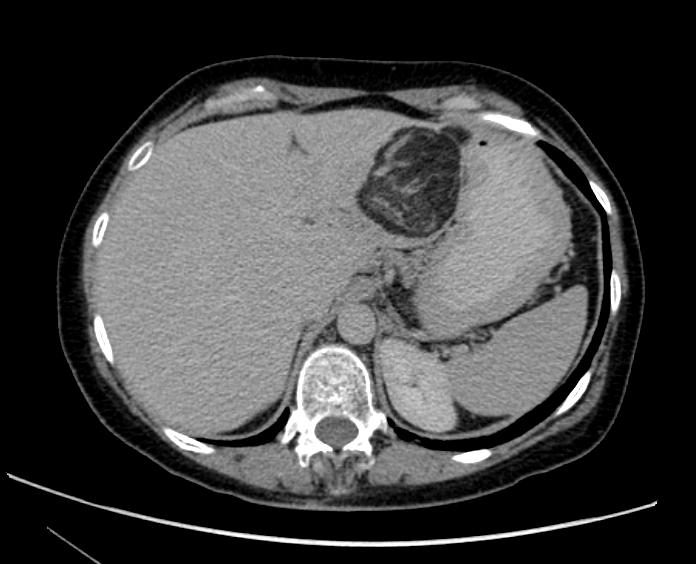
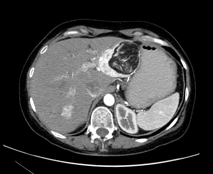
US and CT images demonstrate a large liver angiomyolipoma
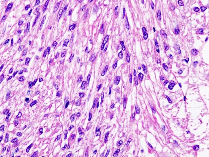
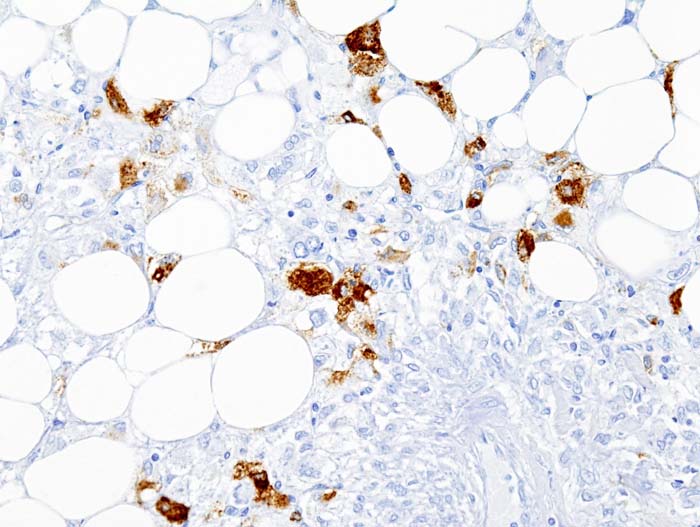
Histopathological Findings
Complications
Complications include hematuria, flank pain, and shock as a result of spontaneous hemorrhage.
References
See also
External links
