Ventricular tachycardia electrocardiogram
|
Ventricular tachycardia Microchapters |
|
Differentiating Ventricular Tachycardia from other Disorders |
|---|
|
Diagnosis |
|
Treatment |
|
Case Studies |
|
Ventricular tachycardia electrocardiogram On the Web |
|
to Hospitals Treating Ventricular tachycardia electrocardiogram |
|
Risk calculators and risk factors for Ventricular tachycardia electrocardiogram |
Editor-In-Chief: C. Michael Gibson, M.S., M.D. [1]; Associate Editor-in Chief: Sara Zand, M.D.[2] Avirup Guha, M.B.B.S.[3]; Priyamvada Singh, M.D. [4]
Overview
Finding on ECG associated with VT include: AV dissociation, atypical right bundle branch block or left bundle branch block characteristics, QRS> 140 ms for wide complex tachycardia with right bundle branch block pattern and QRS > 160 ms for wide complex tachycardia with left bundle branch block pattern, concordance or same polarity in all precordioal leads, rightward superior QRS axis.
Electrocardiogram
Common ECG criteria associated with VT include:[1]
- The key diagnostic criterion for VT especially when the ventricular rate exceeds the atrial rate
- The absence of AV dissociation does not rule out VT
- The series of QRS complexes uncoupled from dissociated P waves
- Limiting the atrial rhythm by self‐governing ventricular rhythm
- Capture beat or single QRS complex resembling the patient's baseline rhythm due to stopping ventricular depolarization by supraventricular impulse
- Fusion beat or a hybrid QRS complex resembling the ventricular depolarization characteristics of the VT and baseline rhythm
- If ventricular impulses conduct retrograde through the His‐Purkinje system to depolarize the atria, VT will not exhibit atrioventricular dissociation.
- Morphologic criteria
- VT is the most likely diagnosis if a wide QRS tachycardia demonstrates a QRS patten incompatible with typical right or left bundle branch block characteristics.
- In the presence of wide QRS tachycardia with atypical right bundle block characteristics including monophasic R wave in V1 or V2 and QS pattern in V6, VT is the most likely diagnosis.
- When there is wide complex tachycardia with classic left bundle branch block pattern ( r wave onset to S wave nadir <60 ms in V1 or V2 and notched monophasic R wave in V6), supraventricular tachycardia is the most likely diagnosis.
- QRS duration
- QRS >140 ms for wide complex tachycardia with right bundle branch block pattern and QRS >160 ms for wide complex tachycardia with left bundle branch block pattern indicating ventricular tachycardia.
- QRS >160 ms may also be seen in supraventricular tachycardia especially among patients with ongoing antiarrhythmic use, electrolyte disturbances, conduction delays, or severe underlying structural heart disease or cardiomyopathies.
- Fascicular VT may demonstrate substantial impulse propagation within the conduction system with QRS durations <120 ms
- Chest Lead Concordance
- QRS complexes in all 6 precordial leads (V1–V6) uniformly shown a monophasic pattern with same polarity ( R for positive concordance and QS for negative concordance)
- Wide complex tachycardias with positive concordance demonstrating VT originating from the posterobasal left ventricle.
- Wide complex tachycardias with negative concordance may arise from VT originating for the anteroapical left ventricle
- Absence of concordance does not rule out VT diagnosis.
- QRS Axis
- Rightward superior QRS axis ( northwest axis) between −90° and −180°
- Dominant R wave in lead avR
- Coexistence of left‐ or right‐axis deviation with right or left bundle branch block
- In the presence of scar‐related VT mapped to the anterolateral wall of the left ventricle may show a wide complex tachycardia with an atypical right bundle branch block pattern and rightward and superior QRS axix which is uncommon in supraventricular tachycardia with right bundle branch block aberrancy.
- Differences in Ventricular Activation Velocity
- slurred initial components of the QRS complex due to slower cardiomyocyte‐to‐cardiomyocyte conduction ( R wave peak time in lead II ≥50 ms, or RS interval ≥100 ms in any of the precordial leads [V1–V6])
- Rapid propagates from conduction system and activation the remainder of the myocardium
- Rapid or sharper deflections in the terminal portion of QRS complex ( the ratio of the voltage excursion during the initial [Vi] and terminal [Vt] 40 ms of the QRS complex <1)
- Comparison to the baseline ECG
- Findings the changes in the QRS axis, T axis, and QRS duration between wide complex tachycardia and baseline ECG ( an ECG taken before or after tachycardia maybe helpful for diagnosis of VT.
| Limb leads algorithm | |||||||||||||||||||||||||||||||||||||||||||||||||||||||
RWPT algorithm
| The VT score
| ||||||||||||||||||||||||||||||||||||||||||||||||||||||
Brugada algorithm
| Ventricular tachycardia algorithm | Vereckie avR algorithm
| |||||||||||||||||||||||||||||||||||||||||||||||||||||
Pachon scoring algorithm
| |||||||||||||||||||||||||||||||||||||||||||||||||||||||
EKG Examples
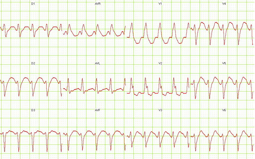
Shown below is an EKG with a rapid ventricular rate of nearly 190 beats per minute with wide QRS complex in all leads depicting ventricular tachycardia.
Copyleft image obtained courtesy of ECGpedia,http://en.ecgpedia.org/wiki/Main_Page
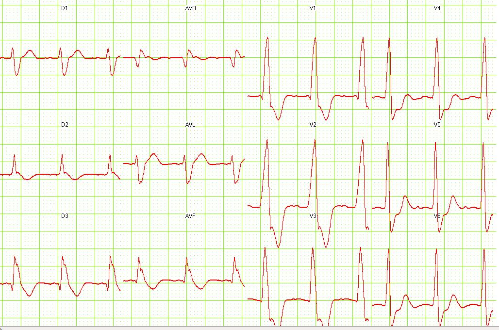
Shown below is an EKG with a rapid ventricular rate of nearly 150 beats per minute with wide QRS complex in all leads depicting ventricular tachycardia.
Copyleft image obtained courtesy of ECGpedia,http://en.ecgpedia.org/wiki/Main_Page
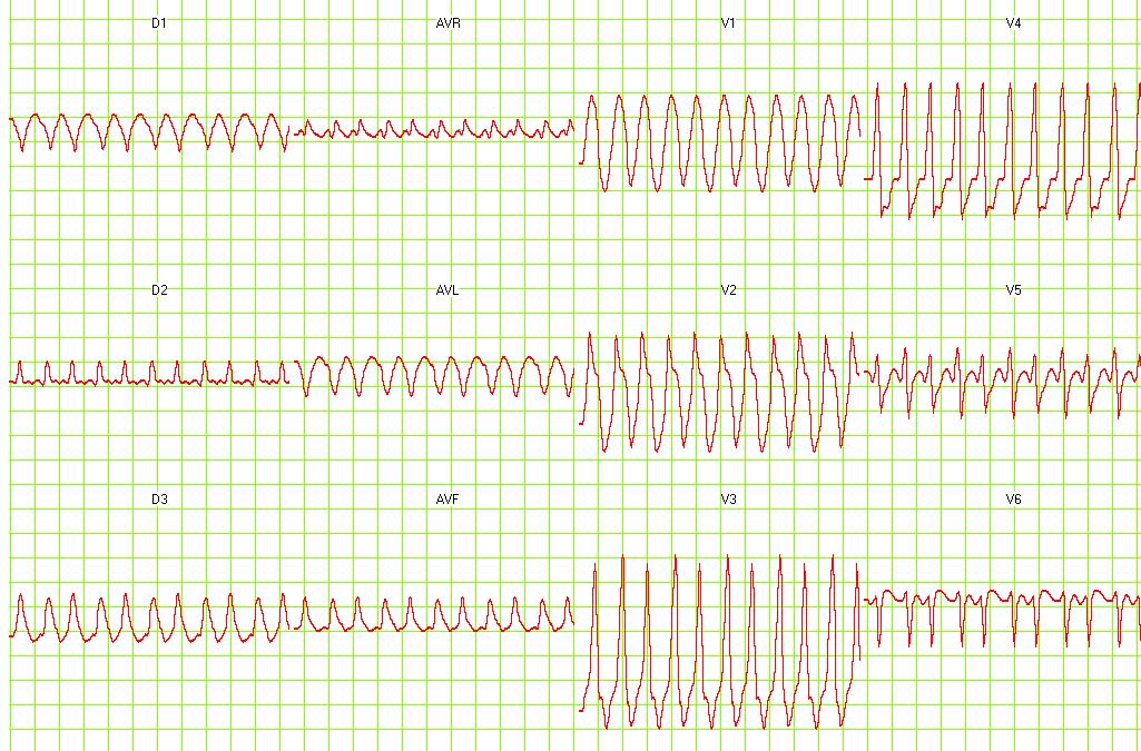
Shown below is an EKG with a rapid ventricular rate of nearly 250 beats per minute with wide QRS complexes in all leads depicting ventricular tachycardia.
Copyleft image obtained courtesy of ECGpedia,http://en.ecgpedia.org/wiki/Main_Page
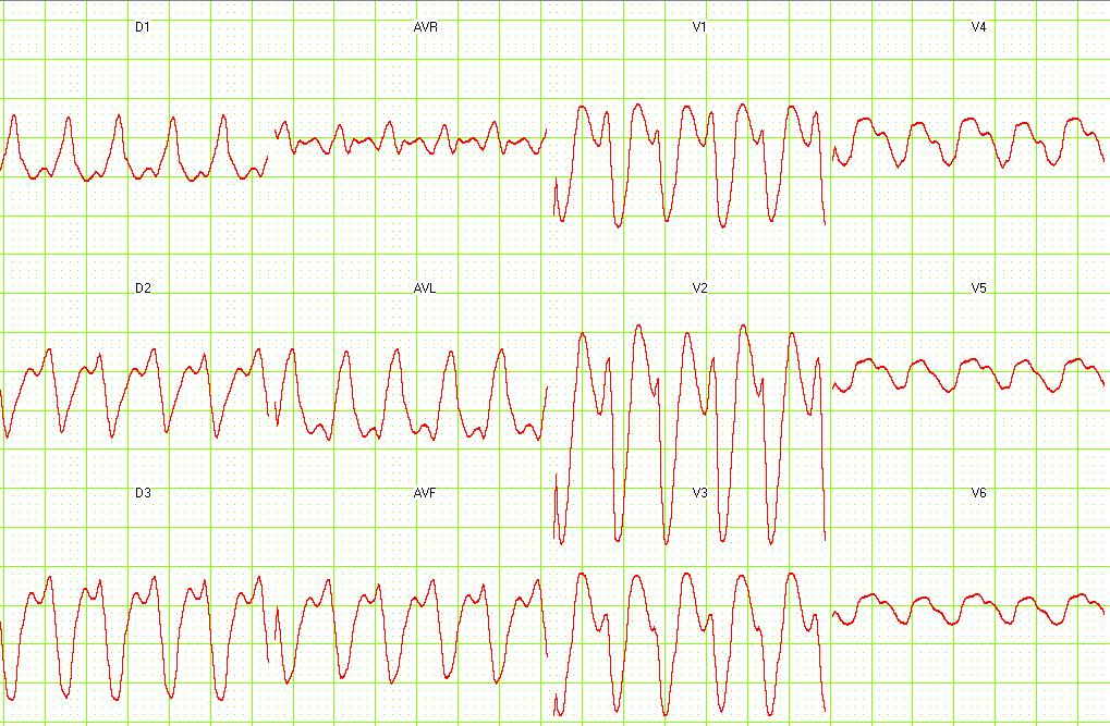
Shown below is an EKG with a rapid ventricular rate of nearly 215 beats per minute with wide QRS complexes in all leads depicting ventricular tachycardia.
Copyleft image obtained courtesy of ECGpedia,http://en.ecgpedia.org/wiki/Main_Page
Shown below is an EKG with a rapid ventricular rate of nearly 140 bpm with a left bundle branch block pattern and left heart axis.
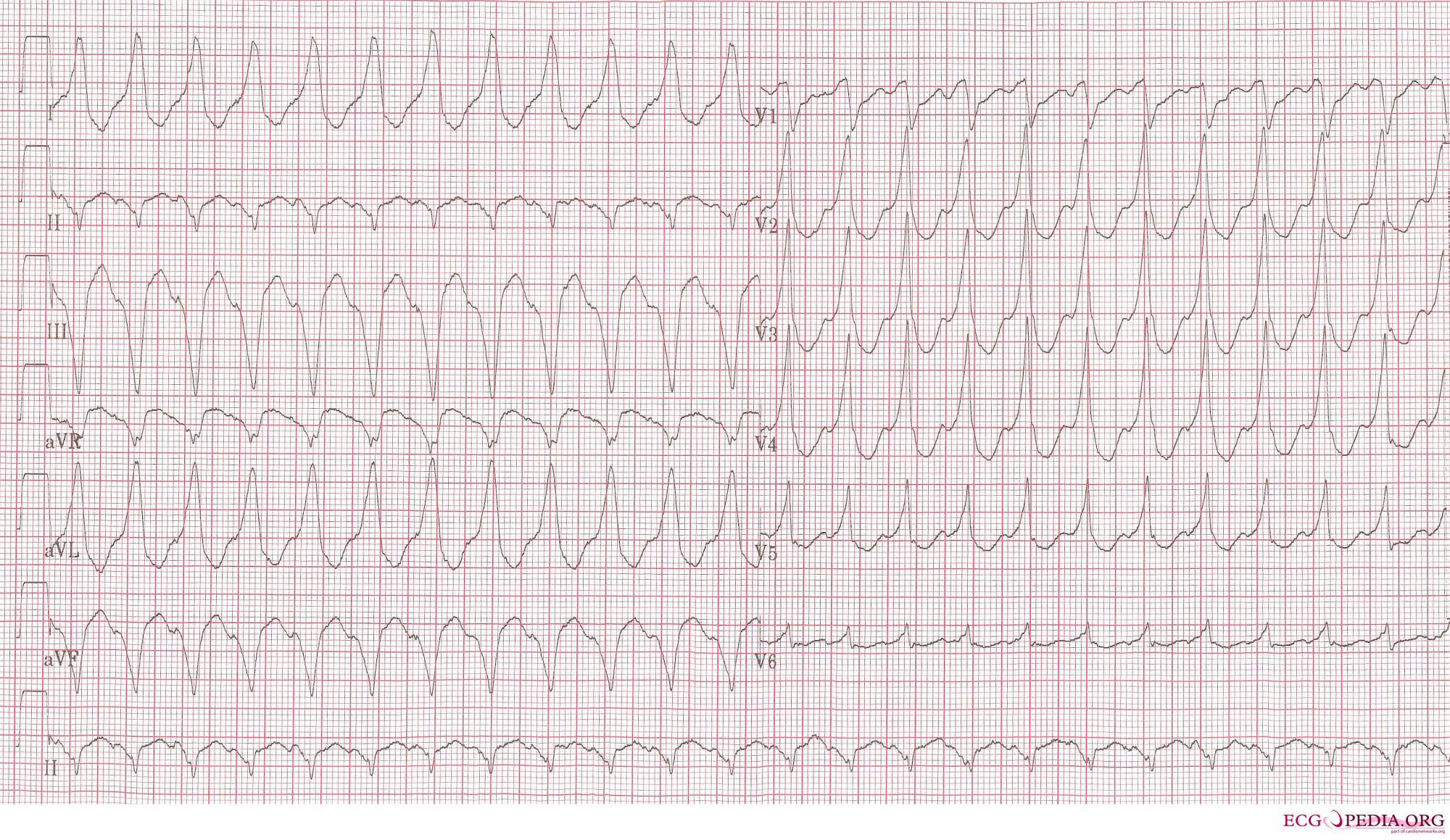
Copyleft image obtained courtesy of ECGpedia,http://en.ecgpedia.org/wiki/Main_Page
Shown below is an EKG depicting ventricular tachycardia with a rate of 250 bpm, and a right bundle branch block pattern with a right heart axis.
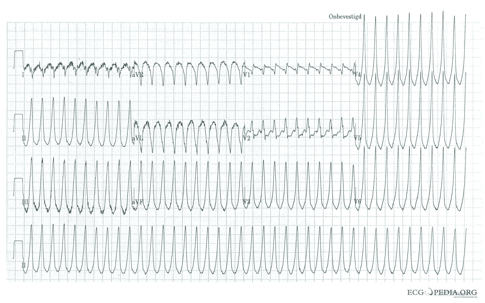
Copyleft image obtained courtesy of ECGpedia,http://en.ecgpedia.org/wiki/Main_Page
Shown below is an EKG depicting ventricular tachycardia with a rate of 150 bpm, and a right bundle branch block pattern with right heart axis. The 5th and 6th complexes from the right side are fusion complexes. Furthermore this EKG shows baseline drift, which is a technical artefact
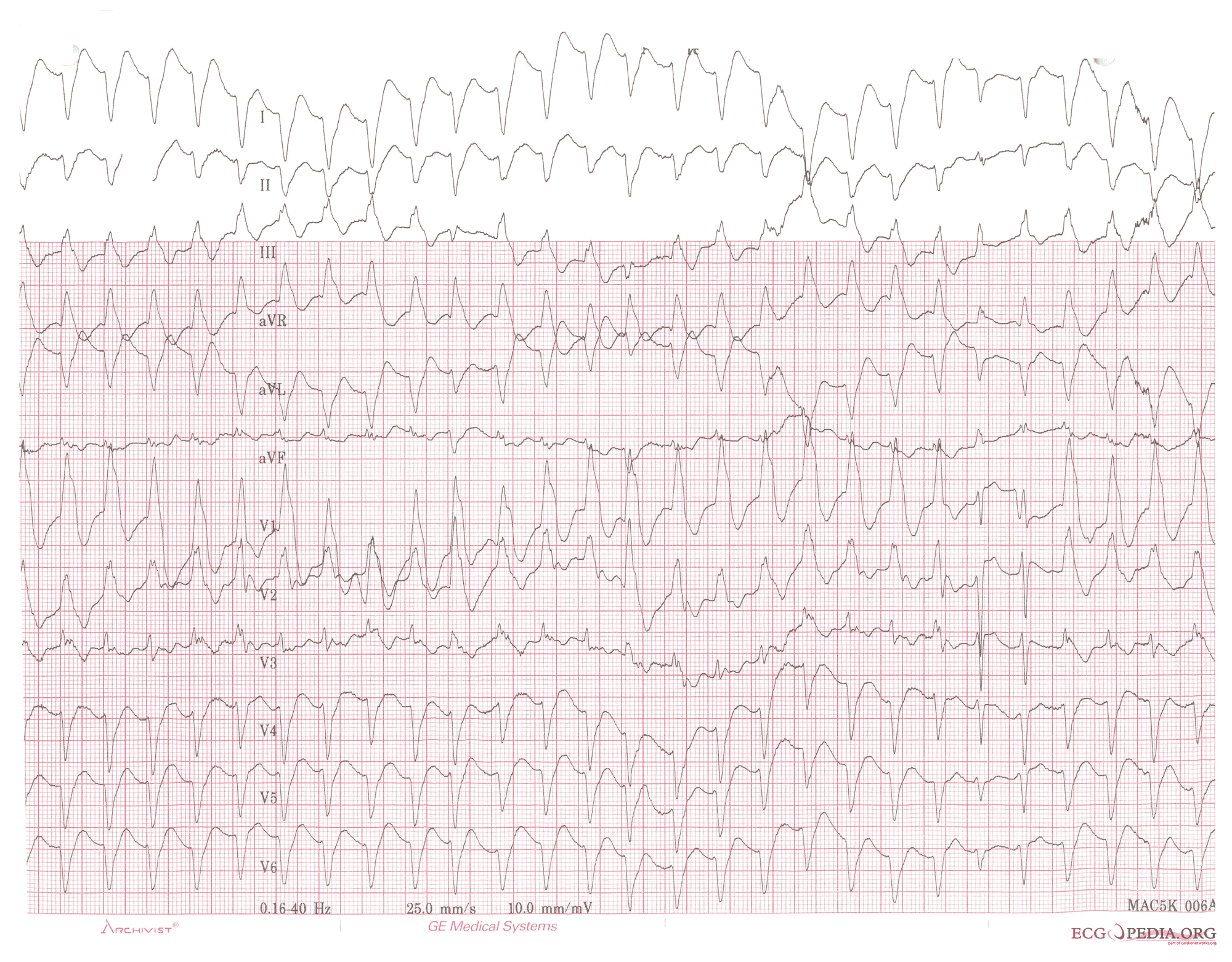
Copyleft image obtained courtesy of ECGpedia,http://en.ecgpedia.org/wiki/Main_Page
Shown below is an EKG depicting a nonsustained VT of five beats duration.
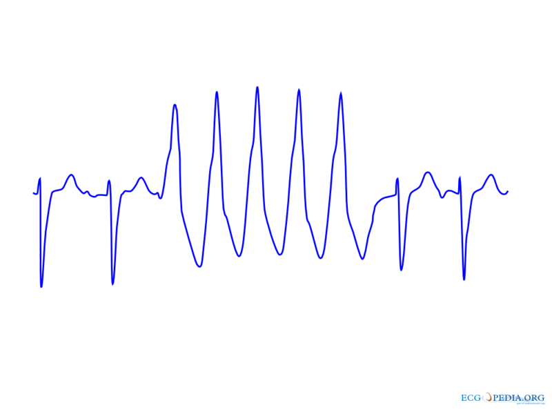
Copyleft image obtained courtesy of ECGpedia,http://en.ecgpedia.org/wiki/File:De-Nsvt.png
Shown below is an EKG depicting ventricular tachycardia at a rate of 145 beats per minute with a right bundle branch block pattern and left heart axis.
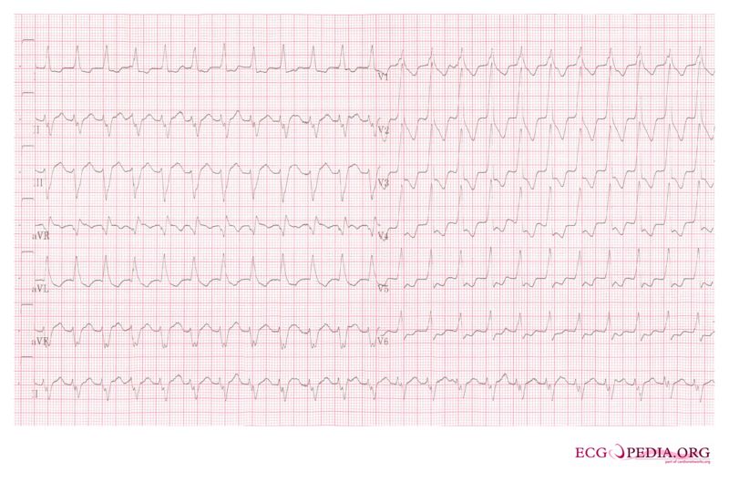
Copyleft image obtained courtesy of ECGpedia,http://en.ecgpedia.org/wiki/File:De-12lead_vt4.jpg
Shown below is an EKG depicting biphasic ventricular tachycardia in a patient with long QT syndrome.
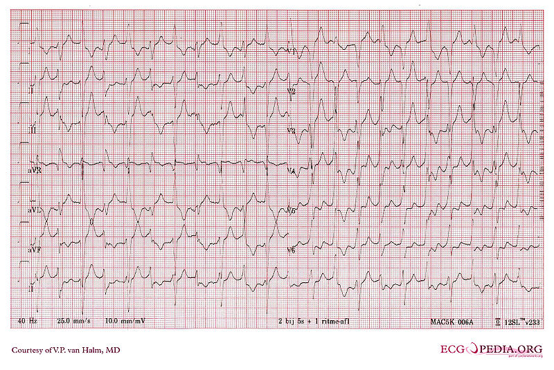
Copyleft image obtained courtesy of ECGpedia,http://en.ecgpedia.org/wiki/File:De-DVA2161.jpg
Shown below is an EKG in a person with idiopathic ventricular tachycardia (Belhassen VT).
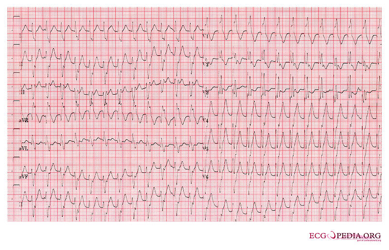
Copyleft image obtained courtesy of ECGpedia,http://en.ecgpedia.org/wiki/File:De-ECG000006.jpg
Shown below is an EKG with a rapid ventricular rate of about 170/min with wide QRS complexes in lead II depicting ventricular tachycardia.

Copyleft image obtained courtesy of ECGpedia, http://en.ecgpedia.org/wiki/Main_Page
Shown below is an EKG depicting a wide complex tachycardia with a left bundle branch morphology at a rate of about 160/min. The R wave in lead V2 is broad, and the time from the beginning of the QRS in lead V2 to the peak of the S wave is longer than 80 ms. No P wave activity is clearly seen. This EKG suggests ventricular tachycardia.
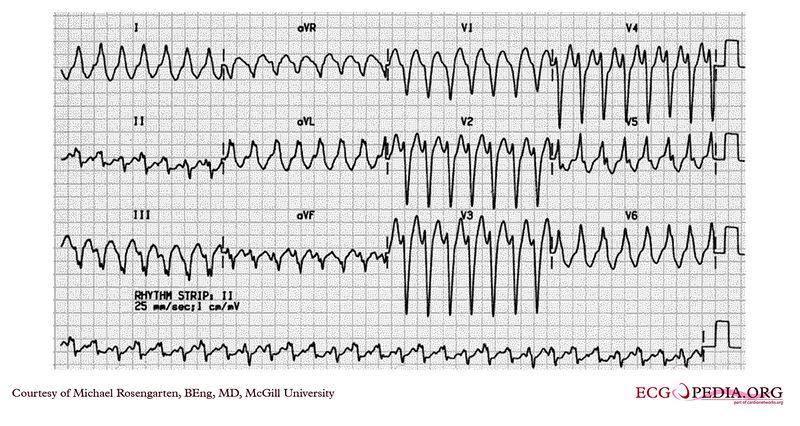
Copyleft image obtained courtesy of ECGpedia,http://en.ecgpedia.org/wiki/File:E334.jpg
Shown below is an EKG with a rapid ventricular rate of nearly 190 beats per minute with wide QRS complexes depicting ventricular tachycardia.
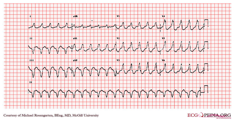
Copyleft image obtained courtesy of ECGpedia,http://en.ecgpedia.org/wiki/File:E253.jpg
Shown below is an EKG with a rapid ventricular rate of about 190/min with wide QRS complex in all leads depicting ventricular tachycardia.
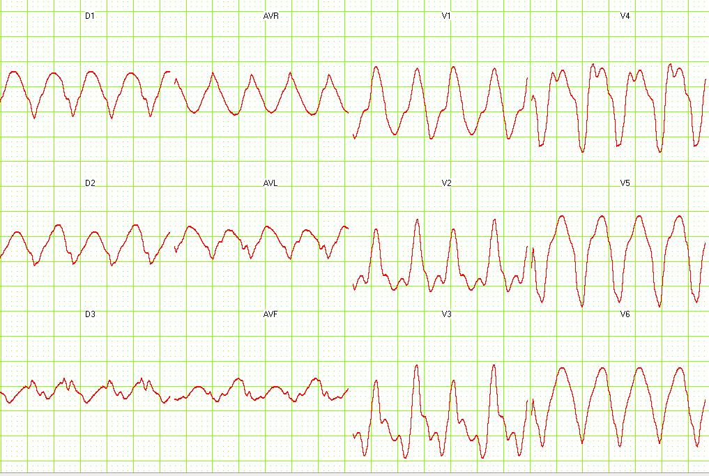
Copyleft image obtained courtesy of ECGpedia, http://en.ecgpedia.org/wiki/Main_Page
For more EKG examples of ventricular tachycardia, click here.
References
- ↑ Kashou, Anthony H.; Noseworthy, Peter A.; DeSimone, Christopher V.; Deshmukh, Abhishek J.; Asirvatham, Samuel J.; May, Adam M. (2020). "Wide Complex Tachycardia Differentiation: A Reappraisal of the State‐of‐the‐Art". Journal of the American Heart Association. 9 (11). doi:10.1161/JAHA.120.016598. ISSN 2047-9980.