Tentorium cerebelli
Editor-In-Chief: C. Michael Gibson, M.S., M.D. [1]
Overview
The tentorium cerebelli or cerebellar tentorium (Latin: "tent of the cerebellum") is an extension of the dura mater that separates the cerebellum from the inferior portion of the occipital lobes.
The tentorium cerebelli is an arched lamina, elevated in the middle, and inclining downward toward the circumference.
It covers the superior surface of the cerebellum, and supports the occipital lobes of the brain.
Its anterior border is free and concave, and bounds a large oval opening, the incisura tentorii, for the transmission of the cerebral peduncles.
It is attached, behind, by its convex border, to the transverse ridges upon the inner surface of the occipital bone, and there encloses the transverse sinuses; in front, to the superior angle of the petrous part of the temporal bone on either side, enclosing the superior petrosal sinuses.
At the apex of the petrous part of the temporal bone the free and attached borders meet, and, crossing one another, are continued forward to be fixed to the anterior and posterior clinoid processes respectively.
To the middle line of its upper surface the posterior border of the falx cerebri is attached, the straight sinus being placed at their line of junction.
Clinically, the tentorium is important because brain tumors are often characterized as supratentorial (above the tentorium) and infratentorial (below the tentorium). The location of the tumor can help in determining the type of tumor, as different tumors occur with different frequencies at each location. Additionally, most childhood tumors are infratentorial, while most adult tumors are supratentorial. The location of the tumor may have prognostic significance as well.
Additional images
-
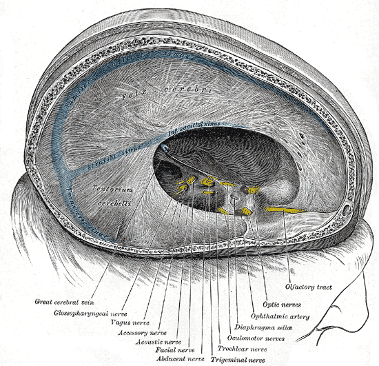 Dura mater and its processes exposed by removing part of the right half of the skull, and the brain.
 -
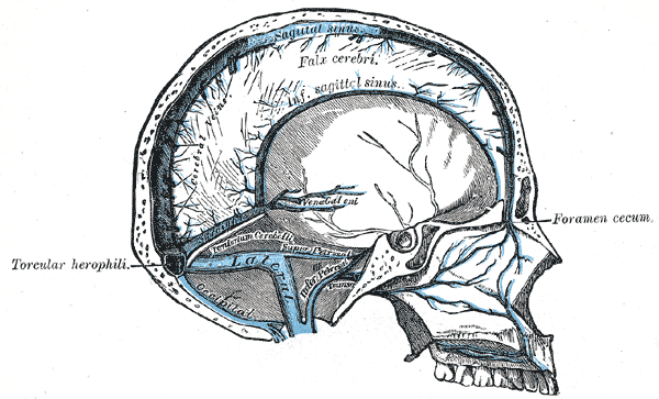 Sagittal section of the skull, showing the sinuses of the dura.

