Extraocular muscles
Editor-In-Chief: C. Michael Gibson, M.S., M.D. [1]

The extraocular muscles are the six muscles that control the movements of the eye. The actions of the extraocular muscles depend on the position of the eye at the time of muscle contraction.
List of muscles
| Muscle | Innervation | Origin | Insertion | Primary function | Secondary function | Tertiary function |
|---|---|---|---|---|---|---|
| Superior rectus | Superior branch of oculomotor nerve | Annulus of Zinn | eye (anterior, superior surface) | Elevation | Intorsion | Adduction |
| Inferior rectus | Inferior branch of oculomotor nerve | Annulus of Zinn | eye (anterior, inferior surface) | Depression | Extorsion | Adduction |
| Lateral rectus | Abducens nerve | Annulus of Zinn | eye (anterior, lateral surface) | Abduction | ||
| Medial rectus | Inferior branch of oculomotor nerve | Annulus of Zinn | eye (anterior, medial surface) | Adduction | ||
| Superior oblique | Trochlear nerve | Annulus of Zinn | eye (posterior, superior, lateral surface) | Intorsion | Depression | Abduction |
| Inferior oblique | Inferior branch of oculomotor nerve | Maxillary bone | eye (posterior, inferior, lateral surface) | Extorsion | Elevation | Abduction |
Actions
| Medial (towards nose) | Lateral (towards temple) |
| Elevation, adduction: Superior rectus |
Elevation, abduction: inferior oblique |
| Adduction: Medial rectus |
Abduction: Lateral rectus |
| Depression, adduction: Inferior rectus |
Depression, abduction: Superior oblique |
In an eye examination, the inability of the patient to move the eye in the specified direction can indicate a problem with the associated muscle, and the nerve associated with that muscle.
Paths

1 = Annulus tendineus communis
2 = Superior rectus muscle
3 = Inferior rectus muscle
4 = Medial rectus muscle
5 = Lateral rectus muscle
6 = Superior oblique muscle
7 = Trochlea of superior oblique
8 = Inferior oblique muscle
9 = Levator palpebrae superioris muscle
10 = Eyelid
11 = Eyeball
12 = Optic nerve
Five with paths from annulus of zinn
Five of the extraocular muscles have their origin in the back of the orbit in a fibrous ring called the annulus of Zinn.
Four of these then course forward through the orbit and insert onto the globe on its anterior half (i.e., in front of the eye's equator). These muscles are named after their straight paths, and are called the four rectus muscles, or four recti.
- superior rectus - inserts on the globe at 2
- inferior rectus - inserts on the globe at 3
- medial rectus - inserts on the globe at 4
- lateral rectus - inserts on the globe at 5
(Note that lateral and medial are relative to the subject, with lateral toward the side and medial toward the midline, thus the medial rectus is the muscle closest to the nose).
Two with more complex paths
The other two extraocular muscles follow more complicated paths.
- The superior oblique muscle originates at the back of the orbit and courses forward to a rigid pulley, called the trochlea, on the upper, nasal wall of the orbit. The muscle passes through the pulley, turning sharply across the orbit, and inserts on the lateral, posterior part of the globe. Thus, the superior oblique goes backward for the last part of its path, and because it goes over the top of the eye, it pulls it downward and nasally (medially).
- The last muscle is the inferior oblique, which originates at the lower front of the nasal orbital wall, and passes under the LR to insert on the lateral, posterior part of the globe. Thus, the inferior oblique pulls the eye upward and nasally (medially).[1][2]
Mnemonic
A good mnemonic to remember which muscles are innervated by what nerve is to paraphrase it as a molecular equation: LR6SO4R3.[3]
- Lateral Rectus - Cranial Nerve VI
- Superior Oblique - Cranial Nerve IV
- the Rest of the muscles - Cranial Nerve III.
Another way to remember which nerves innervate which muscles is to understand the meaning behind all of the Latin words.
- The fourth cranial nerve, the trochlear, is so named because the muscle it innervates, the superior oblique, runs through a little fascial pulley that changes its direction of pull (the trochlea of superior oblique). This pulley exists in the superiomedial corner of each orbit, and "trochl-" is Latin for "pulley."
- The sixth cranial nerve, the abducens, is so named because it controls the lateral rectus, which abducts the eye (rotates it laterally) upon contraction.
- The third cranial nerve, the oculomotor, is so named because it is in charge of the movement (motor) of the eye (oculo-). It controls all of the other muscles.
See also
Additional images
-
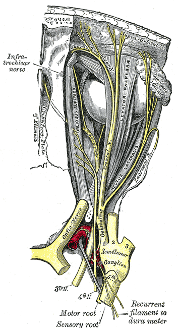 Nerves of the orbit. Seen from above.
 -
 Figure showing the mode of innervation of the Recti medialis and lateralis of the eye.
 -
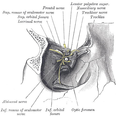 Dissection showing origins of right ocular muscles, and nerves entering by the superior orbital fissure.


References
- ↑ Roger H.S. Carpenter (1988); Movements of the Eyes (2nd ed.). Pion Ltd, London. ISBN 0-85086-109-8.
- ↑ Westheimer Gerald, McKee Suzanne P (1975); "Visual acuity in the presence of retinal-image motion". Journal of the Optical Society of America 65(7), 847-50.
- ↑ {{Medicalmnemonics.com|572|||}}
External links
- neuro/637 at eMedicine - "Extraocular Muscles, Actions"
- Template:UMichAtlas