Left ventricle
|
WikiDoc Resources for Left ventricle |
|
Articles |
|---|
|
Most recent articles on Left ventricle Most cited articles on Left ventricle |
|
Media |
|
Powerpoint slides on Left ventricle |
|
Evidence Based Medicine |
|
Clinical Trials |
|
Ongoing Trials on Left ventricle at Clinical Trials.gov Trial results on Left ventricle Clinical Trials on Left ventricle at Google
|
|
Guidelines / Policies / Govt |
|
US National Guidelines Clearinghouse on Left ventricle NICE Guidance on Left ventricle
|
|
Books |
|
News |
|
Commentary |
|
Definitions |
|
Patient Resources / Community |
|
Patient resources on Left ventricle Discussion groups on Left ventricle Patient Handouts on Left ventricle Directions to Hospitals Treating Left ventricle Risk calculators and risk factors for Left ventricle
|
|
Healthcare Provider Resources |
|
Causes & Risk Factors for Left ventricle |
|
Continuing Medical Education (CME) |
|
International |
|
|
|
Business |
|
Experimental / Informatics |
Editor-In-Chief: C. Michael Gibson, M.S., M.D. [1]
Overview
The left ventricle is one of four chambers (two atria and two ventricles) in the human heart. It receives oxygenated blood from the left atrium via the mitral valve, and pumps it into the aorta via the aortic valve.
Shape
The left ventricle is longer and more conical in shape than the right, and on transverse section its concavity presents an oval or nearly circular outline. It forms a small part of the sternocostal surface and a considerable part of the diaphragmatic surface of the heart; it also forms the apex of the heart.
Development
By teenage and adult ages, its walls have thickened to three to six times greater than that of the right ventricle. This reflects the typical five times greater pressure workload this chamber performs while accepting blood returning from the lungs veins at ~8mmHg pressure and pushing it forward to the typical ~120mmHg pressure in the aorta during each heartbeat. (The pressures stated are resting values and stated as relative to surrounding atmospheric which is the typical "0" reference pressure used in medicine.)
Function
For excellence of health, the left ventricular muscle must:
- (a) relax very rapidly after each contraction so as to fill rapidly with oxygenated blood flowing from the lung veins, i.e. diastolic relaxation and filling.
- (b) contract rapidly and forcibly to force the majority of this blood into the aorta, overcoming the much higher aortic pressure and the extra pressure required to stretch the aorta and other major arteries enough to expand and make room for the sudden increase in blood volume, i.e. systolic contraction and ejection.
- (c) be able to rapidly increase or decrease its pumping capacity under nervous system control.
Pumping volume
Typical healthy adult heart pumping volume is ~5 liters/min, resting. Maximum capacity pumping volume extends from ~25 liters/min for non-athletes to as high as ~45 liters/min for Olympic level athletes.
Additional images
-
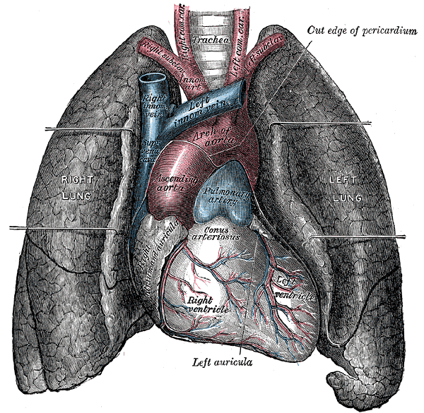 Front view of heart and lungs.
 -
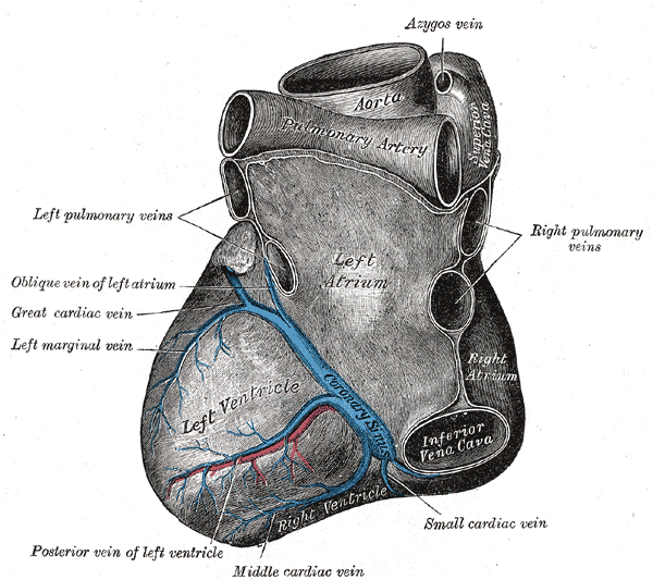 Base and diaphragmatic surface of heart.

