Human parainfluenza virus
|
Croup Microchapters |
|
Diagnosis |
|---|
|
Treatment |
|
Case Studies |
|
Human parainfluenza virus On the Web |
|
American Roentgen Ray Society Images of Human parainfluenza virus |
|
Risk calculators and risk factors for Human parainfluenza virus |
| Human parainfluenza virus | ||||||
|---|---|---|---|---|---|---|
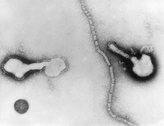
![A Transmission Electron Micrograph (TEM) depicting parainfluenza virions, and free filamentous nucleocapsid material.[1]](/index.php/File:HPIV04.jpeg) A Transmission Electron Micrograph (TEM) depicting parainfluenza virions, and free filamentous nucleocapsid material.[1]
| ||||||
| Virus classification | ||||||
|
Editor-In-Chief: C. Michael Gibson, M.S., M.D. [1] Associate Editor(s)-in-Chief: Luke Rusowicz-Orazem, B.S.
Overview
Human parainfluenza virus is an enveloped, single stranded negative sense RNA virus with four distinct serotypes. The virus genome consists of approximately 15,000 nucleotides used to encode six structural proteins; they function to attach, enter, and fuse with the host cell, forming a complex with the RNA genome. Human parainfluenza virus is a member of the paramyxoviridae family. It is a member of one of two genuses depending on the serotype: Respirovirus or Rubulavirus. Human parainfluenza virus infects the body by infiltrating white blood cells. It is transmitted through respiratory droplets through the air, as well as physical contact with an infected individual or contaminated physical surface.
Microbiological Characteristics
- The human parainfluenza virus genome consists of approximately 15,000 nucleotides used to encode the following six structural proteins:.[4]
| Protein | Location | Function |
|---|---|---|
| hemagglutinin | Envelope | Attachment and cell entry |
| fusion protein | Envelope | Fusion and cell entry |
| matrix protein | Within the envelope | Assembly |
| nucleoprotein | Nucleocapsid | Forms a complex with the RNA genome |
| phosphoprotein | Nucleocapsid | Forms as a part of the RNA polymerase complex |
| large protein | Nucleocapsid | Forms as a part of the RNA polymerase complex |
- Human parainfluenza virus is a member of the paramyxoviridae family.
- The genus for human parainfluenza virus depends on its serotype:[4]
- Respirovirus: HPIV-1 & HPIV-3
- Rubulavirus: HPIV-2 & HPIV-4
Transmission
- Human parainfluenza virus is primarily transmitted by the following:[5]
Virology
- Human parainfluenza virus infiltrates histiocytes, lymphocytes, plasma cells, and neutrophils white blood cells.[6].
- HPIV fuses with the white blood cells through the glycoproteins hemagglutinin-neuraminidase and fusion protein.[4]
- Upon fusion, the HPIV nucleocapsid is expelled into the recipient cell cytoplasm.[4]
- Viral transcription occurs through virus-specific RNA-dependent RNA polymerase.[4]
- The viral mRNAs are translated into viral proteins, leading to the replication of the genome into the following:[4]
- The negative-sense RNA strand is encapsidated by nucleoprotein and is then used for further transcription and replication.
References
- ↑ "phil.cdc.gov".
- ↑ Vainionpää R, Hyypiä T (1994). "Biology of parainfluenza viruses". Clin. Microbiol. Rev. 7 (2): 265–75. PMC 358320. PMID 8055470.
- ↑ Baron, Samuel (1996). Medical microbiology. Galveston, Tex: University of Texas Medical Branch at Galveston. ISBN 0-9631172-1-1.
- ↑ 4.0 4.1 4.2 4.3 4.4 4.5 Henrickson, K. J. (2003). "Parainfluenza Viruses". Clinical Microbiology Reviews. 16 (2): 242–264. doi:10.1128/CMR.16.2.242-264.2003. ISSN 0893-8512.
- ↑ "Human Parainfluenza Viruses | Transmission of HPIVs | CDC".
- ↑ Cherry, James D. (2008). "Croup". New England Journal of Medicine. 358 (4): 384–391. doi:10.1056/NEJMcp072022. ISSN 0028-4793.