Duodenal atresia pathophysiology
|
Duodenal Atresia Microchapters |
|
Diagnosis |
|---|
|
Treatment |
|
Case Studies |
|
Duodenal atresia pathophysiology On the Web |
|
American Roentgen Ray Society Images of Duodenal atresia pathophysiology |
|
Risk calculators and risk factors for Duodenal atresia pathophysiology |
Editor-In-Chief: C. Michael Gibson, M.S., M.D. [1]; Associate Editor(s)-in-Chief: Hamid Qazi, MD, BSc [2]
Overview
It is thought that duodenal atresia is the result of failure of neural cell migration during the 8th to 10th week of duodenal re-canalization. It is associated with down syndrome, vertebral defects, anal anomalies, esophageal atresia, annular pancreas, malrotation, renal abnormalities, cardiac causes, and mandibulofacial anomalies.
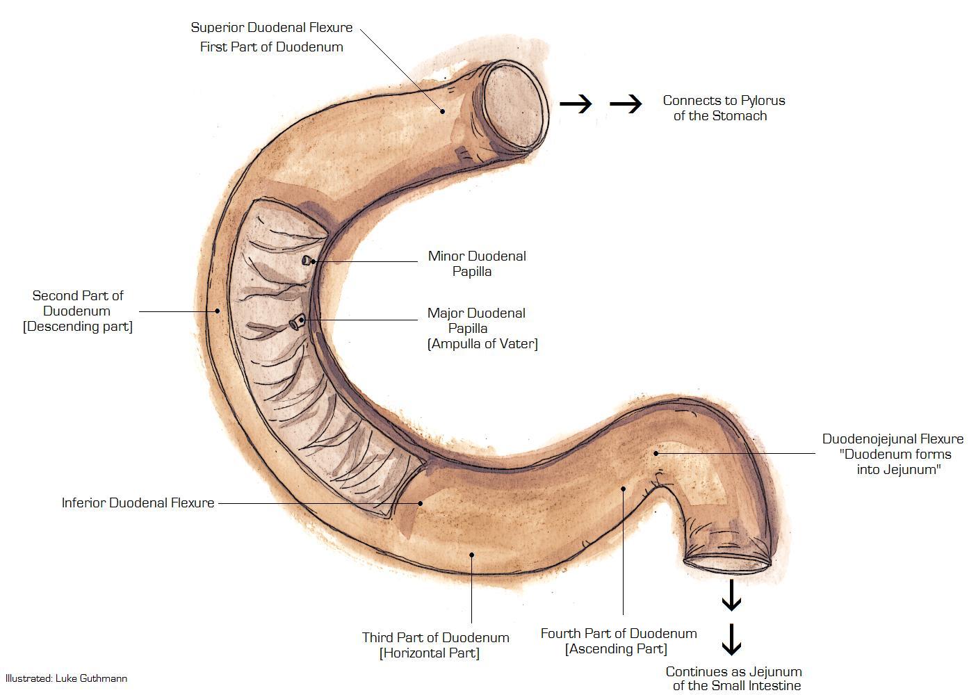
Pathophysiology
Pathogenesis
- Duodenum starts developing during the 6th and 7th week of gestation.[1][2]
- Re-canalization occurs during the 8th to 10th week of gestation.
- It is thought that duodenal atresia is the result of failure of re-canalization of the duodenum in 8 to 10 weeks of fetal development.
- This is due to failure of neural cell migration

Genetics
- Duodenal atresia is not transmitted genetically.
Associated Conditions
Duodenal atresia is commonly associated with the following:[3][4]
- Down syndrome in 25 %to 40% of cases
- VATER
- Vertebral defects
- Anal anomalies
- Esophageal atresia
- Renal abnormalities
- Malrotation
- Annular pancreas
- Biliary tract abnormalities
- Cardiac anomalies
- Mandibulofacial anomalies
References
- ↑ Ando H, Kaneko K, Ito F, Seo T, Harada T, Watanabe Y (1999). "Embryogenesis of pancreaticobiliary maljunction inferred from development of duodenal atresia". J Hepatobiliary Pancreat Surg. 6 (1): 50–4. PMID 10436237.
- ↑ Boyden EA, Cope JG, Bill AH (1967). "Anatomy and embryology of congenital intrinsic obstruction of the duodenum". Am J Surg. 114 (2): 190–202. PMID 6028984.
- ↑ Freeman, SB; Torfs, CP; Romitti, PA; Royle, MH; Druschel, C; Hobbs, CA; Sherman, SL (2009). "Congenital gastrointestinal defects in Down syndrome: a report from the Atlanta and National Down Syndrome Projects". Clinical Genetics. 75 (2): 180–184. doi:10.1111/j.1399-0004.2008.01110.x. ISSN 0009-9163.
- ↑ Morris, Grant; Kennedy, Alfred; Cochran, William (2016). "Small Bowel Congenital Anomalies: a Review and Update". Current Gastroenterology Reports. 18 (4). doi:10.1007/s11894-016-0490-4. ISSN 1522-8037.