Chromosome
| Chromosome | |
 | |
|---|---|
| A representation of a condensed eukaryotic chromosome, as seen during cell division |
|
WikiDoc Resources for Chromosome |
|
Articles |
|---|
|
Most recent articles on Chromosome |
|
Media |
|
Evidence Based Medicine |
|
Clinical Trials |
|
Ongoing Trials on Chromosome at Clinical Trials.gov Clinical Trials on Chromosome at Google
|
|
Guidelines / Policies / Govt |
|
US National Guidelines Clearinghouse on Chromosome
|
|
Books |
|
News |
|
Commentary |
|
Definitions |
|
Patient Resources / Community |
|
Patient resources on Chromosome Discussion groups on Chromosome Patient Handouts on Chromosome Directions to Hospitals Treating Chromosome Risk calculators and risk factors for Chromosome
|
|
Healthcare Provider Resources |
|
Causes & Risk Factors for Chromosome |
|
Continuing Medical Education (CME) |
|
International |
|
|
|
Business |
|
Experimental / Informatics |
Editor-In-Chief: C. Michael Gibson, M.S., M.D. [1]
Associate Editor-In-Chief: Cafer Zorkun, M.D., Ph.D. [2]
Overview
A chromosome is a single large macromolecule of DNA, and constitutes a physically organized form of DNA in a cell. It is a very long, continuous piece of DNA (a single DNA molecule), which contains many genes, regulatory elements and other intervening nucleotide sequences.
A broader definition of "chromosome" also includes the DNA-bound proteins which serve to package and manage the DNA. The word chromosome comes from the Greek Template:Polytonic (chroma, color) and Template:Polytonic (soma, body) due to its capacity to be stained very strongly with vital and supravital dyes.
Chromosomes vary extensively between different organisms. The DNA molecule may be circular or linear, and can contain anything from tens of kilobase pairs to hundreds of megabase pairs.
Typically eukaryotic cells (cells with nuclei) have large linear chromosomes and prokaryotic cells (cells without nuclei) smaller circular chromosomes, although there are many exceptions to this rule. Furthermore, cells may contain more than one type of chromosome; for example mitochondria in most eukaryotes and chloroplasts in plants have their own small chromosome in addition to the nuclear chromosomes.
In eukaryotes nuclear chromosomes are packaged by proteins (particularly histones) into chromatin to fit the massive molecules into the nucleus.
The structure of chromatin varies through the cell cycle, and is responsible for the compaction of DNA into the classic four-arm structure during mitosis and meiosis. Prokaryotes do not form chromatin, because the cells lack proteins required and the circular configuration of the molecule prevents this.
"Chromosome" is a rather loosely defined term. In prokaryotes, a small circular DNA molecule may be called either a plasmid or a small chromosome. In viruses, mitochondria, and chloroplasts their DNA molecules are commonly referred to as chromosomes, despite being naked molecules, as they constitute the complete genome of the organism or organelle.
History
Chromosomes were first observed in plant cells by a Swiss botanist named Karl Wilhelm von Nägeli in 1842, and independently in Ascaris worms by Belgian scientist Edouard Van Beneden (1846-1910).
The use of basophilic aniline dyes was a fundamentally new technique for effectively staining the chromatin material in the nucleus. Their behavior in animal (salamander) cells was later described in detail by German cytologist and professor of anatomy Walther Flemming, the discoverer of mitosis, in 1882.
The name was invented later by another German anatomist, Heinrich von Waldeyer in 1888.
Chromosomes in eukaryotes
Eukaryotes (cells with nuclei such as plants, yeast, and animals) possess multiple large linear chromosomes contained in the cell's nucleus. Each chromosome has one centromere, with one or two arms projecting from the centromere, although under most circumstances these arms are not visible as such.
In addition most eukaryotes have a small circular mitochondrial genome, and some eukaryotes may have additional small circular or linear cytoplasmic chromosomes.
In the nuclear chromosomes of eukaryotes, the uncondensed DNA exists in a semi-ordered structure, where it is wrapped around histones (structural proteins), forming a composite material called chromatin.
-
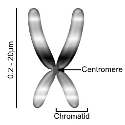
Figure 1: A representation of a condensed eukaryotic chromosome, as seen during cell division.
Chromatin
Chromatin is the complex of DNA and protein found in the eukaryotic nucleus which packages chromosomes. The structure of chromatin varies significantly between different stages of the cell cycle, according to the requirements of the DNA.
-
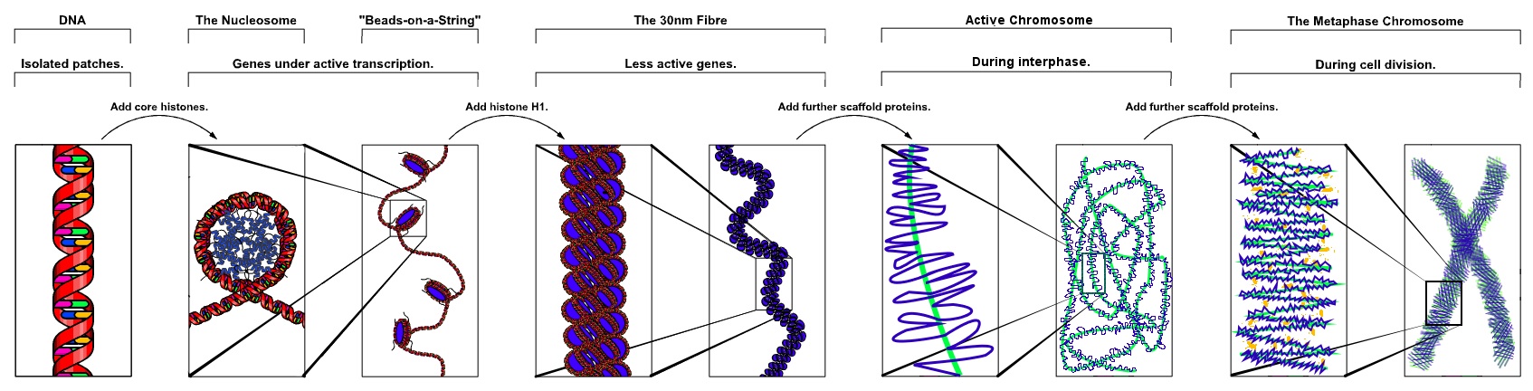
Fig. 2: The major structures in DNA compaction; DNA, the nucleosome, the 10nm "beads-on-a-string" fibre, the 30nm fibre and the metaphase chromosome.

Interphase chromatin
During interphase (the period of the cell cycle where the cell is not dividing) two types of chromatin can be distinguished:
- Euchromatin, which consists of DNA that is active, e.g., expressed as protein.
- Heterochromatin, which consists of mostly inactive DNA. It seems to serve structural purposes during the chromosomal stages. Heterochromatin can be further distinguished into two types:
- Constitutive heterochromatin, which is never expressed. It is located around the centromere and usually contains repetitive sequences.
- Facultative heterochromatin, which is sometimes expressed.
Individual chromosomes cannot be distinguished at this stage - they appear in the nucleus as a homogeneous tangled mix of DNA and protein.
Metaphase chromatin and division
In the early stages of mitosis or meiosis (cell division), the chromatin strands become more and more condensed. They cease to function as accessible genetic material (transcription stops) and become a compact transportable form. This compact form makes the individual chromosomes visible, and they form the classic four arm structure, a pair of sister chromatids attached to each other at the centromere. The shorter arms are called p arms (from the French petit, small) and the longer arms are called q arms (q follows p in the Latin alphabet). This is the only natural context in which individual chromosomes are visible with an optical microscope.
During divisions long microtubules attach to the centromere and the two opposite ends of the cell. The microtubules then pull the chromatids apart, so that each daughter cell inherits one set of chromatids. Once the cells have divided, the chromatids are uncoiled and can function again as chromatin. In spite of their appearance, chromosomes are structurally highly condensed which enables these giant DNA structures to be contained within a cell nucleus (Fig. 2).
The self assembled microtubules form the spindle, which attaches to chromosomes at specialized structures called kinetochores, one of which is present on each sister chromatid. A special DNA base sequence in the region of the kinetochores provides, along with special proteins, longer-lasting attachment in this region.
-
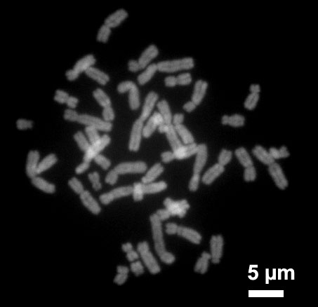
Human chromosomes during metaphase.

Chromosomes in prokaryotes
Prokaryotes (eg. Bacteria) typically have a single circular chromosome, but many variations do exist.
Bacterial DNA also exists as plasmids, essentially miniature chromosomes, which are small circular pieces of DNA that are readily transmitted between bacteria. The distinction between plasmids and chromosomes is poorly defined, though size and necessity are generally taken into account.
Structure in sequences
Prokaryotes chromosomes have less sequence-based structure than eukaryotes. They do, however, typically have a single point, the origin of replication, from which replication starts.
The genes in prokaryotes are often organised in operons, and do not contain introns, unlike eukaryotes.
Location in the cell
Bacterial chromosomes tend to be tethered to the plasma membrane of the bacteria. In molecular biology application, this allows for its isolation from plasmid DNA by centrifugation of lysed bacteria and pelleting of the membranes (and the attached DNA).
DNA packaging
Prokaryotes do not possess histones or nuclei, and so do not possess chromatin like eukaryotes. There is, however, thought to be some structural organisation to help condense the large molecule into the small prokaryotic cell.
Prokaryotic chromosomes and plasmids are, like eukaryotic DNA, generally supercoiled. The DNA must first be released into its relaxed state for access for transcription, regulation, and replication.
Number of chromosomes in various organisms
Eukaryotes
|
| ||||||||||||||||||||||||||||||||||||||||||||||||||||||||||||||||||||||||||||||||||||||||||||||||||||||||||||
 Normal members of a particular eukaryotic species all have the same number of nuclear chromosomes (see the table). Other eukaryotic chromosomes, i.e., mitochondrial and plasmid-like small chromosomes, are much more variable in number, and there may be thousands of copies per cell. Asexually reproducing species have one set of chromosomes, which is the same in all body cells. Sexually reproducing species have somatic cells (body cells), which are diploid [2n] having two sets of chromosomes, one from the mother and one from the father. Gametes, reproductive cells, are haploid [n]: they have one set of chromosomes. Gametes are produced by meiosis of a diploid germ line cell. During meiosis, the matching chromosomes of father and mother can exchange small parts of themselves (crossover), and thus create new chromosomes that are not inherited solely from either parent. When a male and a female gamete merge (fertilization), a new diploid organism is formed. Some animal and plant species are polyploid [Xn]: they have more than two sets of homologous chromosomes. Agriculturally important plants such as tobacco or wheat are often polyploid compared to their ancestral species. Wheat has a haploid number of seven chromosomes, still seen in some cultivars as well as the wild progenitors. The more common pasta and bread wheats are polyploid, having 28 (tetraploid) and 42 (hexaploid) chromosomes compared to the 14 (diploid) chromosomes in the wild wheat.[9] Historical note: In 1921, Theophilus Painter claimed, based on his observations, that human sex cells had 24 chromosomes each, giving humans 48 chromosomes total. It wasn't until 1955 that the number of chromosomes was clearly shown to be 23. ProkaryotesProkaryote species generally have one copy of each major chromosome, but most cells can easily survive with multiple copies. Plasmids and plasmid-like small chromosomes are, like in eukaryotes, very variable in copy number. The number of plasmids in the cell is almost entirely determined by the rate of division of the plasmid - fast division causes high copy number, and vice versa. Karyotype Karyotyping is a technique used to determine the (diploid) number of nuclear chromosomes of a eukaryotic organism, and may be used for determining sex and spotting chromosomal abnormalities. Cells can be locked part way through division (in metaphase) in vitro (in a reaction vial) with colchicine. These cells are then stained, photographed and arranged into a karyotype (an ordered set of chromosomes, Fig. 3), also called karyogram. Like many sexually reproducing species, humans have special gonosomes (sex chromosomes, in contrast to autosomes). These are XX in females and XY in males, and can be seen in the karyotype, Fig. 3. Chromosomal aberrations   Chromosomal aberrations are disruptions in the normal chromosomal content of a cell, and are a major cause of genetic conditions in humans, such as Down syndrome. Some chromosome abnormalities do not cause disease in carriers, such as translocations, or chromosomal inversions, although they may lead to a higher chance of having a child with a chromosome disorder. Abnormal numbers of chromosomes or chromosome sets, aneuploidy, may be lethal or give rise to genetic disorders. Genetic counseling is offered for families that may carry a chromosome rearrangement. The gain or loss of chromosome material can lead to a variety of genetic disorders. Human examples include:
Chromosomal mutations produce changes in whole chromosomes (more than one gene) or in the number of chromosomes present.
Most mutations are neutral- have little or no effect A detailed graphical display of all human chromosomes and the diseases annotated at the correct spot may be found at [4]. Human chromosomesHuman cells have 23 pairs of large linear nuclear chromosomes, giving a total of 46 per cell. In addition to these, human cells have many hundreds of copies of the mitochondrial genome. Sequencing of the human genome has provided a great deal of information about each of the chromosomes. Below is a table compiling statistics for the chromosomes, based on the Sanger Institute's human genome information in the Vertebrate Genome Annotation (VEGA) database.[10] Number of genes is an estimate as it is in part based on gene predictions. Total chromosome length is an estimate as well, based on the estimated size of unsequenced heterochromatin regions.
References
See also
Structural Diagrams
External links
ar:صبغي bn:ক্রোমোজোম bg:Хромозома ca:Cromosoma cs:Chromozóm cy:Cromosom da:Kromosom de:Chromosom et:Kromosoom eo:Kromosomo fa:رنگینتن gl:Cromosoma ko:염색체 hr:Kromosomi id:Kromosom it:Cromosoma he:כרומוזום ka:ქრომოსომა ku:Kromozom lv:Hromosoma lt:Chromosoma lmo:Cromusoma hu:Kromoszóma mk:Хромозом nl:Chromosoom no:Kromosom simple:Chromosome sk:Chromozóm sr:Хромозом fi:Kromosomi sv:Kromosom th:โครโมโซม ur:لونجسیمہ yi:כראמאזאם |
|||||||||||||||||||||||||||||||||||||||||||||||||||||||||||||||||||||||||||||||||||||||||||||||||||||||||||||


