Cardiac tamponade CT scan
Jump to navigation
Jump to search
|
Cardiac tamponade Microchapters |
|
Diagnosis |
|---|
|
Treatment |
|
Case Studies |
|
Cardiac tamponade CT scan On the Web |
|
American Roentgen Ray Society Images of Cardiac tamponade CT scan |
|
Risk calculators and risk factors for Cardiac tamponade CT scan |
Editor-In-Chief: C. Michael Gibson, M.S., M.D. [1]; Associate Editor(s)-in-Chief: Ramyar Ghandriz MD[2]
Overview
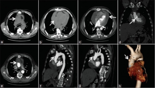
A CT scan is not commonly used for the diagnosis of cardiac tamponade as it is effectively diagnosed based on clinical features and echocardiography. Findings on CT include [Superior vena cava]] andInferior vena cava enlargement, Hepatic and renal vein enlargment, periportal edema, reflux of contrast material, collapse of the right atrium, Pericardial thickening.
CT scan
- CT scan provides pivotal information about the underlying cause of pericardial effusion based on attenuation measurements of collection.[1][2][3][4][5]
- The following are the findings on CT scan:
- Superior vena cava andInferior vena cava enlargement
- Hepatic and renal vein enlargment
- Periportal edema
- Reflux of contrast material
- Compression of coronary sinus
- Angulation of interventricular septum
- Pericardial thickening
- Collapse of the right atrium
- Aortic blood contrast level

References
- ↑ "Cardiac tamponade | Radiology Reference Article | Radiopaedia.org".
- ↑ Xu B, Harb SC, Klein AL (March 2018). "Utility of multimodality cardiac imaging in disorders of the pericardium". Echo Res Pract. doi:10.1530/ERP-18-0019. PMC 5911773. PMID 29588309.
- ↑ Hoey ET, Shahid M, Watkin RW (June 2016). "Computed tomography and magnetic resonance imaging evaluation of pericardial disease". Quant Imaging Med Surg. 6 (3): 274–84. doi:10.21037/qims.2016.01.03. PMC 4929280. PMID 27429911.
- ↑ Baxi AJ, Restrepo C, Mumbower A, McCarthy M, Rashmi K (November 2015). "Cardiac Injuries: A Review of Multidetector Computed Tomography Findings". Trauma Mon. 20 (4): e19086. doi:10.5812/traumamon.19086. PMC 4727463. PMID 26839855.
- ↑ Ünal E, Karcaaltincaba M, Akpinar E, Ariyurek OM (March 2019). "The imaging appearances of various pericardial disorders". Insights Imaging. 10 (1): 42. doi:10.1186/s13244-019-0728-4. PMC 6441059. PMID 30927107.
- ↑ Shergill, Arvind K; Maraj, Tishan; Barszczyk, Mark S; Cheung, Helen; Singh, Navneet; Zavodni, Anna E (2015). "Identification of Cardiac and Aortic Injuries in Trauma with Multi-detector Computed Tomography". Journal of Clinical Imaging Science. 5: 48. doi:10.4103/2156-7514.163992. ISSN 2156-7514.