Widened mediastinum
| Widened mediastinum | |
 | |
|---|---|
| Widened mediastinum |
Editor-In-Chief: C. Michael Gibson, M.S., M.D. [1]; Associate Editor-In-Chief: Varun Kumar, M.B.B.S.
Overview
A widened mediastinum is a mediastinum which measures greater than 8 cm in width on PA chest X-ray. A widened mediastinum can be indicative of life threatening conditions such as aortic dissection and esophageal rupture.
Anatomy
- Anterior mediastinum: Extends from the sternum back to the anterior line of the heart. Contains the thymus, fat, and lymph nodes.
- Middle mediastinum: Contains the heart, pericardium, ascending and transverse aorta, brachiocephalic vessels, superior vena cava, inferior vena cava, main pulmonary arteries and veins, trachea, bronchi and lymph nodes.
- Posterior mediastinum: From the posterior border of the heart and trachea to the anterior surface of the thoracic spine and ribs. Contains the descending aorta, the esophagus, the azygous vein, autonomic ganglia and nerves, thoracic duct, lymph nodes, and fat.
Causes
Common Causes
- Anterior mediastinal mass
- Teratoma
- Lymphomas - Hodgkins and non-hodgkins. Anterosuperior, smooth and homogenous, and usually surrounds great vessels.
- Germ cell tumor - may contain calcifications.
- Thymoma and other thymic abnormalities (thymic carcinomas, thymic cysts, thymolipomas).
- Thyroid (intrathoracic goiter) - thoracic inlet, smooth and frequently symmetric.
- Connective tissue tumors
- Middle mediastinal mass
- Pericardial cyst - usually right sided
- Bronchogenic cyst - frequently subcarinal
- Enteric cyst
- Lymphadenopathy- from infectious, malignant and idiopathic etiologies.
- Posterior mediastinal mass
- Neurogenic tumor
- Esophageal tumors
- Neuroenteric cysts
- Hiatal hernia
Causes by Organ System
| Cardiovascular | |
| Chemical / poisoning | No underlying causes |
| Dermatologic | Dermoid cyst |
| Drug Side Effect | No underlying causes |
| Ear Nose Throat |
|
| Endocrine |
|
| Environmental | Intrathoracic goitre |
| Gastroenterologic |
|
| Genetic | No underlying causes |
| Hematologic | |
| Iatrogenic | Mediastinitis |
| Infectious Disease |
|
| Musculoskeletal / Ortho | No underlying causes |
| Neurologic | Mediastinal neurilemmoma may originate from right phrenic nerve[9], intrathoracic vagal nerve[10] |
| Nutritional / Metabolic | Intrathoracic goitre |
| Obstetric/Gynecologic | No underlying causes |
| Oncologic | |
| Opthalmologic | No underlying causes |
| Overdose / Toxicity | Inhaled recreational drugs such as cocaine which induced bronschospasm, increased alveolar pressure followed by alveolar rupture leading to interstitial emphysema and pneumomediastinum[11]. |
| Psychiatric | No underlying causes |
| Pulmonary |
|
| Renal / Electrolyte | No underlying causes |
| Rheum / Immune / Allergy | |
| Sexual | No underlying causes |
| Trauma |
|
| Urologic | No underlying causes |
| Miscellaneous |
|
Causes in Alphabetical Order
- Anthrax
- This is a classic finding associated with inhalational anthrax. A widened mediastinum was found in 7 of the first 10 victims infected by anthrax (Bacillus anthracis) in 2001.[8]
- Aortic dissection
- Bronchogenic cyst
- Churg-Strauss syndrome
- Dermoid cyst
- Esophageal achalasia
- Esophageal cancer
- Esophageal rupture
- Goitre
- Hiatus hernia
- Hilar lymphadenopathy
- Lymphoma
- Mediastinal germ cell tumor
- Mediastinal tumor
- Mediastinal mass
- Mediastinitis
- Neurilemmoma
- Non-Hodgkin lymphoma
- Partial anomalous pulmonary venous connection
- Pericardial effusion
- Pneumomediastinum
- Sarcoidosis
- Superior vena cava obstruction
- Supine AP chest x ray can yield a false positive "widened mediastinum".
- Among patients who have sustained blunt traum, AP chest radiographs are often obtained in the supine position to maintain spinal precautions. This supine position may result in fluid shifts that are in turn associated with a widening of the mediastinum. After the spine had been "cleared", some authors recommend repeating the chest X ray with the patient in the erect position which results in normalization of the mediastinal size in around 40% of patients.
- Teratoma
- Thymoma
- Thyroid cancer
- Tularemia
Diagnosis
Due to an extensive differential diagnosis list for a widened mediastinum, life-threatening situations such as aortic dissection and esophageal rupture should be ruled out first. If a mass is visualized on a plain radiograph, its specific location should be determined and further visualization using CT with contrast can aid in the specific diagnosis. Laboratory testing for infectious, rheumatologic, hematologic, and endocrine abnormalities, based on a thorough history and physical examination and imaging can lead to diagnosis in the remaining causes of a widened mediastinum.
Diagnosis in the Acute Setting
Widening of the mediastinum on the chest X ray may represent a medical emergency and the following diagnoses should be excluded immediately:
- Aortic dissection
- Esophageal rupture (Boerhaave's syndrome)
To confirm a widened mediastinum, a measurement of more than or equal to 8cm at the level of the aortic knob on a good quality plain film should be obtained.In ruling out the presence of traumatic aortic injury in a patient, a widened mediastinum is reported as having a 53% sensitivity, 59% specificity and 83% negative predictive value.
In the evaluation of aortic dissection or aortic injury, other less sensitive signs on the chest x ray include the following:
- Depression of the left main-stem bronchus
- Deviation of the trachea or the esophagus to the right
- Apical pleural hematoma (also known as a pleural cap)
- Disruption of the calcium ring in the aortic knob (broken-halo)
- First and second rib fracture
- Obliterated aorto-pulmonary window
Multi-detector spiral CT scan has widely replaced the use of angiography for the initial evaluation of a widened mediastinum in the setting of trauma. Chest CT should be obtained even if there is a negative chest radiograph in the setting of trauma, due to the false negative appearance of a normal mediastinum despite aortic injury being present. If no periarotic mediastinal blood can be visualized on CT, then aortic injury can be ruled out. Trans-esophageal echocardiography is also a good method of imaging which has a sensitivity and specificity approaching 100%.
In the setting of a blunt deceleration trauma, disruption of the thoracic aorta can occur. This injury usually occurs at the ductus arteriosum just distal to the take-off of the left subclavian artery. Occasionally, the aorta may rupture in the ascending portion and at the take-off of the major vessels. If aortic dissection is suspected, asses for symptomatic blood pressure differences (of more that 10mmHg), pseudocoarctation syndrome or infrascapular murmur.
Esophageal rupture usually occurs after esophageal surgery due to injury with a surgical instrument, but can also occur due to trauma or repeated vomiting, and can be spontaneous. Initial evaluation is with a chest radiograph which will show a widened mediastinum with free air in the peritoneum. This should be followed up with contrast studies of the esophagus, and a CT scan.
Chest X Ray
Shown below is the development of a new widened mediastinum in a patient with aortic dissection:
-
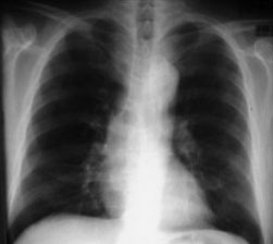
Chest x ray of a patient 3 months before an aortic dissection
-
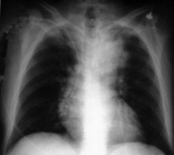
Chest x ray of the same patient the day of admission for aortic dissection showing a new widened mediastinum


Diagnosis in the Setting of a Mediastinal Mass
To narrow the differential of a mediastinal mass in the setting of a widened mediastinum, it is useful to know in what part of the mediastinal cavity the mass is located. A mediastinal mass is first detected and evaluated on a chest radiograph, and then further visualized with a chest CT scan, preferably with intravenous water-soluble contrast material. MRI rarely adds any additional imaging information for most mediastinal masses, however it is indicated for the evaluation of neurogenic tumors, and may also provide additional information in the evaluation of cystic lesions and vascular invasion. MRI is also useful to image a mediastinal mass in a patient with a contrast allergy or renal failure. Ultrasonographic evaluation can be useful to assess for cystic versus solid masses, in particular those that are close to the heart.
The age of the patient can also help in narrowing down the differential in the diagnosis of a mediastinal mass in the setting of a widened mediastinum. In infants and children, it is likely that the mass will be a neurogenic tumor or an enterogenous cyst. In adults, neurogenic tumors, thymomas and thymic cysts are the most common causes. Between the ages of 20 and 40, a malignant mass should be high on the differential, as this is the age range at which Hodgkins and Non-hodgkins lymphoma presents.
References
- ↑ Netterville JL, Coleman SC, Smith JC, Smith MM, Day TA, Burkey BB (1998). "Management of substernal goiter". Laryngoscope. 108 (11 Pt 1): 1611–7. PMID 9818814.
- ↑ MKSAP 11: Medical knowledge self-assessment program. Philadelphia: American College of Physicians, 1998:966-7.
- ↑ Corsten MJ, Shamji FM, Odell PF, Frederico JA, Laframboise GG, Reid KR, et al. Optimal treatment of descending necrotising mediastinitis. Thorax 1997;52:702-8.
- ↑ Wheatley MJ, Stirling MC, Kirsch MM, Gago O, Orringer MB. Descending necrotizing mediastinitis: transcervical drainage is not enough. Ann Thorac Surg 1990;49:780-4.
- ↑ Estrera AS, Landay MJ, Grisham JM, Sinn DP, Platt MR. Descending necrotizing mediastinitis. Surg Gynecol Obstet 1983;157:545-52.
- ↑ Alsoub H, Chacko KC. Descending necrotising mediastinitis. Postgrad Med J 1995;71:98-101.
- ↑ Sakamoto H, Aoki T, Kise Y, Watanabe D, Sasaki J (2000). "Descending necrotizing mediastinitis due to odontogenic infections". Oral Surg Oral Med Oral Pathol Oral Radiol Endod. 89 (4): 412–9. PMID 10760723.
- ↑ 8.0 8.1 Jernigan JA, Stephens DS, Ashford DA; et al. (2001). "Bioterrorism-related inhalational anthrax: the first 10 cases reported in the United States". Emerging Infect. Dis. 7 (6): 933–44. PMID 11747719.
- ↑ Hirose H, Ohmori K, Nakaoka Y, Kitamura K, Muramatsu T, Namiki Y; et al. (1998). "[Mediastinal neurilemmoma originating in the right phrenic nerve: a case report]". Nihon Kokyuki Gakkai Zasshi. 36 (12): 1027–31. PMID 10064956.
- ↑ Ito I, Komota K, Nakajima T, Ishibashi K, Kawazoe K (1994). "[A case of mediastinal neurilemmoma originating from the intrathoracic vagal nerve]". Kyobu Geka. 47 (4): 325–7. PMID 8152184.
- ↑ Panacek EA, Singer AJ, Sherman BW, Prescott A, Rutherford WF (1992). "Spontaneous pneumomediastinum: clinical and natural history". Ann Emerg Med. 21 (10): 1222–7. PMID 1416301.
- ↑ MKSAP 11: Medical knowledge self-assessment program. Philadelphia: American College of Physicians, 1998:966-7.
- ↑ Corsten MJ, Shamji FM, Odell PF, Frederico JA, Laframboise GG, Reid KR, et al. Optimal treatment of descending necrotising mediastinitis. Thorax 1997;52:702-8.
- ↑ Wheatley MJ, Stirling MC, Kirsch MM, Gago O, Orringer MB. Descending necrotizing mediastinitis: transcervical drainage is not enough. Ann Thorac Surg 1990;49:780-4.
- ↑ Estrera AS, Landay MJ, Grisham JM, Sinn DP, Platt MR. Descending necrotizing mediastinitis. Surg Gynecol Obstet 1983;157:545-52.
- ↑ Alsoub H, Chacko KC. Descending necrotising mediastinitis. Postgrad Med J 1995;71:98-101.