Neck of femur fracture classification: Difference between revisions
(Created page with " __NOTOC__ {{Neck of femur fracture}} {{CMG}}; {{AE}} {{Rohan}} ==Overview== There are multiple classifications available for neck of femur fracture. The...") |
m (Bot: Removing from Primary care) |
||
| (3 intermediate revisions by 2 users not shown) | |||
| Line 1: | Line 1: | ||
__NOTOC__ | |||
{{Neck of femur fracture}} | {{Neck of femur fracture}} | ||
| Line 5: | Line 5: | ||
==Overview== | ==Overview== | ||
There are multiple [[Classification|classifications]] available for neck of femur fracture. The most common classification systems for neck of femur fracture include Anatomical, Garden's, Pauwel's and AO/OTA [[classification]]. | There are multiple [[Classification|classifications]] available for neck of femur fracture. The most common classification systems for neck of [[femur fracture]] include Anatomical, Garden's, Pauwel's and AO/OTA [[classification]]. | ||
==Classification== | ==Classification== | ||
| Line 16: | Line 16: | ||
|} | |} | ||
*Anatomical classification is the commonly used classification for | *Anatomical classification is the commonly used classification for eck of femur fracture.<ref>{{cite book | last = Rockwood | first = Charles | title = Rockwood and Green's fractures in adults | publisher = Wolters Kluwer Health/Lippincott Williams & Wilkins | location = Philadelphia, PA | year = 2010 | isbn = 9781605476773 }}</ref><ref>{{cite book | last = Azar | first = Frederick | title = Campbell's operative orthopaedics | publisher = Elsevier | location = Philadelphia, PA | year = 2017 | isbn = 9780323374620 }}</ref> | ||
{| class="wikitable" | {| class="wikitable" | ||
| Line 22: | Line 22: | ||
|- | |- | ||
| style="background: #4479BA; color: #FFFFFF; text-align: center;" |Type I | | style="background: #4479BA; color: #FFFFFF; text-align: center;" |Type I | ||
| style="background: #F5F5F5; padding: 5px; text-align: center;" | | | style="background: #F5F5F5; padding: 5px; text-align: center;" |Supcapital [[Bone fracture|fracture]] | ||
|- | |- | ||
| style="background: #4479BA; color: #FFFFFF; text-align: center;" | Type II | | style="background: #4479BA; color: #FFFFFF; text-align: center;" | Type II | ||
| style="background: #F5F5F5; padding: 5px; text-align: center;" | | | style="background: #F5F5F5; padding: 5px; text-align: center;" |Transcervical [[Bone fracture|fracture]] | ||
|- | |- | ||
| style="background: #4479BA; color: #FFFFFF; text-align: center;" |Type III | | style="background: #4479BA; color: #FFFFFF; text-align: center;" |Type III | ||
| style="background: #F5F5F5; padding: 5px; text-align: center;" | | | style="background: #F5F5F5; padding: 5px; text-align: center;" |Basicervical [[Bone fracture|fracture]] | ||
|} | |} | ||
===Garden's Classification=== | ===Garden's Classification=== | ||
* Garden's [[classification]] of neck of femur fracture is most commonly used [[classification]].<ref> Garden RS. Low-angle fixation in fractures of the femoral neck. J Bone Joint Surg Br 1961;43-B:647-63. </ref> | * Garden's [[classification]] of neck of femur fracture is most commonly used [[classification]].<ref>Garden RS. Low-angle fixation in fractures of the femoral neck. J Bone Joint Surg Br 1961;43-B:647-63. </ref> | ||
* It is based on anterioposterior (AP) radiographs and does not consider lateral or sagittal plane alignment. | |||
{| class="wikitable" | {| class="wikitable" | ||
| Line 47: | Line 39: | ||
|- | |- | ||
| style="background: #4479BA; color: #FFFFFF; text-align: center;" |Type I | | style="background: #4479BA; color: #FFFFFF; text-align: center;" |Type I | ||
| style="background: #F5F5F5; padding: 5px; text-align: center;" | | | style="background: #F5F5F5; padding: 5px; text-align: center;" |Incomplete, valgus impacted [[fracture]] | ||
|- | |- | ||
| style="background: #4479BA; color: #FFFFFF; text-align: center;" |Type II | | style="background: #4479BA; color: #FFFFFF; text-align: center;" |Type II | ||
| style="background: #F5F5F5; padding: 5px; text-align: center;" | | | style="background: #F5F5F5; padding: 5px; text-align: center;" |Complete, nondisplaced [[Bone fracture|fracture]] | ||
|- | |- | ||
| style="background: #4479BA; color: #FFFFFF; text-align: center;" |Type III | | style="background: #4479BA; color: #FFFFFF; text-align: center;" |Type III | ||
| style="background: #F5F5F5; padding: 5px; text-align: center;" | | | style="background: #F5F5F5; padding: 5px; text-align: center;" |Complete, partially displaced [[Bone fracture|fracture]] | ||
|- | |- | ||
| style="background: #4479BA; color: #FFFFFF; text-align: center;" |Type IV | | style="background: #4479BA; color: #FFFFFF; text-align: center;" |Type IV | ||
| style="background: #F5F5F5; padding: 5px; text-align: center;" | | | style="background: #F5F5F5; padding: 5px; text-align: center;" |Complete, fully displaced [[Bone fracture|fracture]] | ||
|} | |} | ||
===Pauwel's Classification=== | ===Pauwel's Classification=== | ||
* Pauwel's [[Classification|classified]] for neck of femur fracture is based on vertical orientation of fracture line.<ref>Pauwels F. Der Schenkelhalsbruch, ein mechanisches problem. Stuttgart: F. Enke; 1935.</ref> | * Pauwel's [[Classification|classified]] for neck of femur fracture is based on vertical orientation of [[fracture]] line.<ref>Pauwels F. Der Schenkelhalsbruch, ein mechanisches problem. Stuttgart: F. Enke; 1935.</ref> | ||
* Pauwel's angle is defined as the angle formed between the line of a fracture of the neck of the femur and the horizontal on an anterioposterior radiograph. | * Pauwel's angle is defined as the angle formed between the line of a [[fracture]] of the [[neck of the femur]] and the horizontal on an anterioposterior [[Radiography|radiograph]]. | ||
* The greater the angle, the more unstable the fracture and thus worse the prognosis. | * The greater the angle, the more unstable the [[fracture]] and thus worse the [[prognosis]]. | ||
{| class="wikitable" | {| class="wikitable" | ||
! colspan="2" style="background: #4479BA; color: #FFFFFF; text-align: center;" |Pauwel's Classification | ! colspan="2" style="background: #4479BA; color: #FFFFFF; text-align: center;" |Pauwel's Classification | ||
|- | |- | ||
| style="background: #4479BA; color: #FFFFFF; text-align: center;" | | | style="background: #4479BA; color: #FFFFFF; text-align: center;" |Type I | ||
| style="background: #F5F5F5; padding: 5px; text-align: center;" | | | style="background: #F5F5F5; padding: 5px; text-align: center;" |< 30 degree from horizontal | ||
|- | |- | ||
| style="background: #4479BA; color: #FFFFFF; text-align: center;" | | | style="background: #4479BA; color: #FFFFFF; text-align: center;" |Type II | ||
| style="background: #F5F5F5; padding: 5px; text-align: center;" | | | style="background: #F5F5F5; padding: 5px; text-align: center;" |30 to 50 degree from horizontal | ||
|- | |- | ||
| style="background: #4479BA; color: #FFFFFF; text-align: center;" | | | style="background: #4479BA; color: #FFFFFF; text-align: center;" |Type III | ||
| style="background: #F5F5F5; padding: 5px; text-align: center;" | | | style="background: #F5F5F5; padding: 5px; text-align: center;" |> 50 degree from horizontal | ||
|} | |} | ||
===OTA System=== | ===OTA System=== | ||
| Line 91: | Line 77: | ||
|- | |- | ||
| rowspan="4" style="background: #4479BA; color: #FFFFFF; text-align: center;" |A | | rowspan="4" style="background: #4479BA; color: #FFFFFF; text-align: center;" |A | ||
| colspan="2" style="background: #DCDCDC; padding: 5px; text-align: center;" | | | colspan="2" style="background: #DCDCDC; padding: 5px; text-align: center;" |[[Femoral]] [[Trochanteric fossa|Trochanteric]] [[Bone fracture|fractures]] | ||
|- | |- | ||
| style="background: #4479BA; color: #FFFFFF; text-align: center;" |A1 | | style="background: #4479BA; color: #FFFFFF; text-align: center;" |A1 | ||
| style="background: #F5F5F5; padding: 5px; text-align: center;" | | | style="background: #F5F5F5; padding: 5px; text-align: center;" |Simple peritrochanteric | ||
|- | |- | ||
| style="background: #4479BA; color: #FFFFFF; text-align: center;" |A2 | | style="background: #4479BA; color: #FFFFFF; text-align: center;" |A2 | ||
| style="background: #F5F5F5; padding: 5px; text-align: center;" | | | style="background: #F5F5F5; padding: 5px; text-align: center;" |Multifragmentary peritrochanteric, lateral wall incompetent (< 20.5 mm) | ||
|- | |- | ||
| style="background: #4479BA; color: #FFFFFF; text-align: center;" |A3 | | style="background: #4479BA; color: #FFFFFF; text-align: center;" |A3 | ||
| style="background: #F5F5F5; padding: 5px; text-align: center;" | | | style="background: #F5F5F5; padding: 5px; text-align: center;" |Intertrochanteric (reverse obliquity) | ||
|- | |- | ||
| rowspan="4" style="background: #4479BA; color: #FFFFFF; text-align: center;" |B | | rowspan="4" style="background: #4479BA; color: #FFFFFF; text-align: center;" |B | ||
| colspan="2" style="background: #DCDCDC; padding: 5px; text-align: center;" | | | colspan="2" style="background: #DCDCDC; padding: 5px; text-align: center;" |[[Femoral]] [[Neck]] [[Bone fracture|fractures]] | ||
|- | |- | ||
| style="background: #4479BA; color: #FFFFFF; text-align: center;" |B1 | | style="background: #4479BA; color: #FFFFFF; text-align: center;" |B1 | ||
| style="background: #F5F5F5; padding: 5px; text-align: center;" | | | style="background: #F5F5F5; padding: 5px; text-align: center;" |Subcapital | ||
|- | |- | ||
| style="background: #4479BA; color: #FFFFFF; text-align: center;" |B2 | | style="background: #4479BA; color: #FFFFFF; text-align: center;" |B2 | ||
| style="background: #F5F5F5; padding: 5px; text-align: center;" | | | style="background: #F5F5F5; padding: 5px; text-align: center;" |Transcervical | ||
|- | |- | ||
| style="background: #4479BA; color: #FFFFFF; text-align: center;" |B3 | | style="background: #4479BA; color: #FFFFFF; text-align: center;" |B3 | ||
| style="background: #F5F5F5; padding: 5px; text-align: center;" | | | style="background: #F5F5F5; padding: 5px; text-align: center;" |Basicervical | ||
|- | |- | ||
| rowspan=" | | rowspan="3" style="background: #4479BA; color: #FFFFFF; text-align: center;" |C | ||
| colspan="2" style="background: #DCDCDC; padding: 5px; text-align: center;" | | | colspan="2" style="background: #DCDCDC; padding: 5px; text-align: center;" |[[Femoral]] [[Head]] [[Bone fracture|fractures]] | ||
|- | |- | ||
| style="background: #4479BA; color: #FFFFFF; text-align: center;" |C1 | | style="background: #4479BA; color: #FFFFFF; text-align: center;" |C1 | ||
| style="background: #F5F5F5; padding: 5px; text-align: center;" |[[ | | style="background: #F5F5F5; padding: 5px; text-align: center;" |[[Split]] [[Fracture (bone)|fracture]] | ||
|- | |- | ||
| style="background: #4479BA; color: #FFFFFF; text-align: center;" |C2 | | style="background: #4479BA; color: #FFFFFF; text-align: center;" |C2 | ||
| style="background: #F5F5F5; padding: 5px; text-align: center;" |[[ | | style="background: #F5F5F5; padding: 5px; text-align: center;" |Depression [[fracture]] | ||
|} | |} | ||
| Line 134: | Line 117: | ||
[[Category:Orthopedics]] | [[Category:Orthopedics]] | ||
[[Category:Orthopedic surgery]] | [[Category:Orthopedic surgery]] | ||
[[Category:Fractures]] | [[Category:Fractures]] | ||
[[Category:Up-To-Date]] | [[Category:Up-To-Date]] | ||
Latest revision as of 22:55, 29 July 2020
|
Neck of femur fracture Microchapters |
|
Diagnosis |
|---|
|
Treatment |
|
Case Studies |
|
Neck of femur fracture classification On the Web |
|
American Roentgen Ray Society Images of Neck of femur fracture classification |
|
Risk calculators and risk factors for Neck of femur fracture classification |
Editor-In-Chief: C. Michael Gibson, M.S., M.D. [1]; Associate Editor(s)-in-Chief: Rohan A. Bhimani, M.B.B.S., D.N.B., M.Ch.[2]
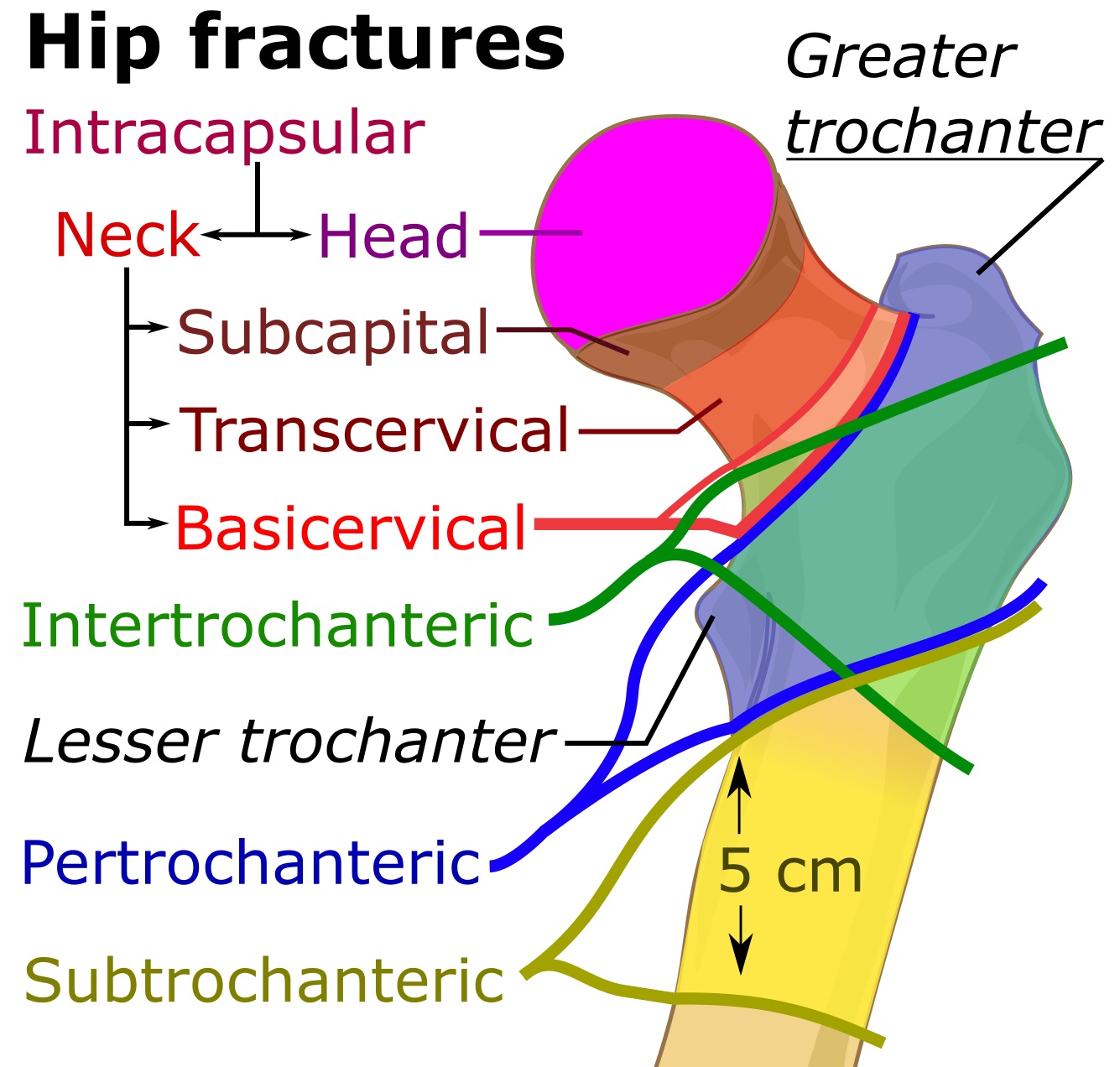
Overview
There are multiple classifications available for neck of femur fracture. The most common classification systems for neck of femur fracture include Anatomical, Garden's, Pauwel's and AO/OTA classification.
Classification
There are multiple classifications available for neck of femur fracture. The most common classification systems for neck of femur fracture include Anatomical, Garden's, Pauwel's and AO/OTA classification.[1][2]
Anatomical Classification
 |
| Schatzker Classification | |
|---|---|
| Type I | Supcapital fracture |
| Type II | Transcervical fracture |
| Type III | Basicervical fracture |
Garden's Classification
- Garden's classification of neck of femur fracture is most commonly used classification.[5]
- It is based on anterioposterior (AP) radiographs and does not consider lateral or sagittal plane alignment.
| Garden's Classification | |
|---|---|
| Type I | Incomplete, valgus impacted fracture |
| Type II | Complete, nondisplaced fracture |
| Type III | Complete, partially displaced fracture |
| Type IV | Complete, fully displaced fracture |
Pauwel's Classification
- Pauwel's classified for neck of femur fracture is based on vertical orientation of fracture line.[6]
- Pauwel's angle is defined as the angle formed between the line of a fracture of the neck of the femur and the horizontal on an anterioposterior radiograph.
- The greater the angle, the more unstable the fracture and thus worse the prognosis.
| Pauwel's Classification | |
|---|---|
| Type I | < 30 degree from horizontal |
| Type II | 30 to 50 degree from horizontal |
| Type III | > 50 degree from horizontal |
OTA System
- AO/ASIF classification is also a widely accepted classification.[7]
- Proximal femur is given the number 31 based on the classification.
- It is further subdivided as:
| OTA System | ||
|---|---|---|
| A | Femoral Trochanteric fractures | |
| A1 | Simple peritrochanteric | |
| A2 | Multifragmentary peritrochanteric, lateral wall incompetent (< 20.5 mm) | |
| A3 | Intertrochanteric (reverse obliquity) | |
| B | Femoral Neck fractures | |
| B1 | Subcapital | |
| B2 | Transcervical | |
| B3 | Basicervical | |
| C | Femoral Head fractures | |
| C1 | Split fracture | |
| C2 | Depression fracture | |
References
- ↑ Rockwood, Charles (2010). Rockwood and Green's fractures in adults. Philadelphia, PA: Wolters Kluwer Health/Lippincott Williams & Wilkins. ISBN 9781605476773.
- ↑ Azar, Frederick (2017). Campbell's operative orthopaedics. Philadelphia, PA: Elsevier. ISBN 9780323374620.
- ↑ Rockwood, Charles (2010). Rockwood and Green's fractures in adults. Philadelphia, PA: Wolters Kluwer Health/Lippincott Williams & Wilkins. ISBN 9781605476773.
- ↑ Azar, Frederick (2017). Campbell's operative orthopaedics. Philadelphia, PA: Elsevier. ISBN 9780323374620.
- ↑ Garden RS. Low-angle fixation in fractures of the femoral neck. J Bone Joint Surg Br 1961;43-B:647-63.
- ↑ Pauwels F. Der Schenkelhalsbruch, ein mechanisches problem. Stuttgart: F. Enke; 1935.
- ↑ ME Muller, S Nazarian, P Koch. Classification AO des fractures. 1 Les os longs. Springler-Verlag, Berlin, 1987.