Endocarditis physical examination: Difference between revisions
| Line 35: | Line 35: | ||
* [[Abdominal pain]] may be present due to mesenteric embolization or [[ileus]] both of which may manifest as reduced bowel sounds | * [[Abdominal pain]] may be present due to mesenteric embolization or [[ileus]] both of which may manifest as reduced bowel sounds | ||
* [[Splenomegaly]] may be present in 15% to 30% patients. | * [[Splenomegaly]] may be present in 15% to 30% patients. | ||
* [[Left upper quadrant pain (LUQ pain) may be present as a result of a [[splenic infarct]] from embolization. | * [[Left upper quadrant pain]] (LUQ pain) may be present as a result of a [[splenic infarct]] from embolization. | ||
* [[Flank pain]] may be present as a result of an [[embolus to the kidney]] | * [[Flank pain]] may be present as a result of an [[embolus to the kidney]] | ||
Revision as of 22:39, 8 October 2012
|
Endocarditis Microchapters |
|
Diagnosis |
|---|
|
Treatment |
|
2014 AHA/ACC Guideline for the Management of Patients With Valvular Heart Disease |
|
Case Studies |
|
Endocarditis physical examination On the Web |
|
Risk calculators and risk factors for Endocarditis physical examination |
Editor-In-Chief: C. Michael Gibson, M.S., M.D. [1]; Associate Editors-in-Chief: Cafer Zorkun, M.D., Ph.D. [2]
Overview
Common signs on physical examination of endocarditis include fever, rigors, Osler's nodes, Janeway lesions and evidence of embolization. Aortic insufficiency with a wide pulse pressure, mitral regurgitation or tricuspid regurgitation may be present depending upon the valve that is infected.
Vital Signs
- A fever will likely be present.
- Rigors may be present.
- Some patients may have a wide pulse pressure due to aortic insufficiency. If the pulse pressure narrows, this may be a sign of left ventricular failure due to earlier closure of the mitral valve and a more rapid rise in the left ventricular end diastolic pressure which will in turn raise the diastolic pressure.
Skin
- Petechiae are present in 10% to 40% of patients
- Splinter hemorrhages are present in 5% to 15% of patients
- Osler's nodes which are tender subcutaneous nodules in pulp of digits are present in 7% to 10% of patients
- Janeway lesions which are erythematous, nontender lesions on palm or sole are present in 6% to 10% of patients
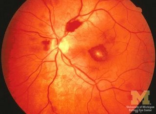
Eyes

Ear Nose and Throat
In patients in whom there is new acute onset of aortic regurgitation, bobbing of the uvula may be present.
Heart
- Heart Murmur(s) are present in 80% to 85% of patients including that of aortic insufficiency, tricuspid regurgitation and mitral regurgitation.
Lungs
- Signs of heart failure such as rales may present
Abdomen
- Abdominal pain may be present due to mesenteric embolization or ileus both of which may manifest as reduced bowel sounds
- Splenomegaly may be present in 15% to 30% patients.
- Left upper quadrant pain (LUQ pain) may be present as a result of a splenic infarct from embolization.
- Flank pain may be present as a result of an embolus to the kidney
Extremities
- Janeway lesions (painless hemorrhagic cutaneous lesions on the palms and soles)
- Gangrene of fingers may occur
- The fingers may show splinter haemorrhages
- Osler's nodes (painful subcutaneous lesions in the distal fingers)
Neurologic
- Septic emboli may result in stroke and focal neurologic findings
- Seizures may be present
- Intracranial hemorrhage may occur
- Signs of a brain abscess may be present