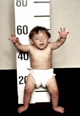
Cyanosis physical examination
|
Cyanosis Microchapters |
|
Diagnosis |
|---|
|
Treatment |
|
Case Studies |
|
Cyanosis physical examination On the Web |
|
American Roentgen Ray Society Images of Cyanosis physical examination |
|
Risk calculators and risk factors for Cyanosis physical examination |
Editor-In-Chief: C. Michael Gibson, M.S., M.D. [1]; Associate Editor(s)-in-Chief: Chandrakala Yannam, MD [2]
Overview
Patients with [disease name] usually appear [general appearance]. Physical examination of patients with [disease name] is usually remarkable for [finding 1], [finding 2], and [finding 3].
OR
Common physical examination findings of [disease name] include [finding 1], [finding 2], and [finding 3]
OR
The presence of [finding(s)] on physical examination is diagnostic of [disease name].
OR
The presence of [finding(s)] on physical examination is highly suggestive of [disease name].
Physical Examination
- Physical examination of patients with [disease name] is usually normal.
OR
- Physical examination of patients with [disease name] is usually remarkable for:[finding 1], [finding 2], and [finding 3].
- The presence of [finding(s)] on physical examination is diagnostic of [disease name].
- The presence of [finding(s)] on physical examination is highly suggestive of [disease name].
Appearance of the Patient
- Patients with [disease name] usually appear [general appearance].
Vital Signs
- High-grade / low-grade fever
- Hypothermia / hyperthermia may be present
- Tachycardia with regular pulse or (ir)regularly irregular pulse
- Bradycardia with regular pulse or (ir)regularly irregular pulse
- Tachypnea / bradypnea
- Kussmal respirations may be present in _____ (advanced disease state)
- Weak/bounding pulse / pulsus alternans / paradoxical pulse / asymmetric pulse
- High/low blood pressure with normal pulse pressure / wide pulse pressure / narrow pulse pressure
Skin
- Skin examination of patients with [disease name] is usually normal.
OR
-
Description (Adapted from Dermatology Atlas)
-
Description (Adapted from Dermatology Atlas)

HEENT
- HEENT examination of patients with [disease name] is usually normal.
OR
- Abnormalities of the head/hair may include ___
- Evidence of trauma
- Icteric sclera
- Nystagmus
- Extra-ocular movements may be abnormal
- Pupils non-reactive to light / non-reactive to accommodation / non-reactive to neither light nor accommodation
- Ophthalmoscopic exam may be abnormal with findings of ___
- Hearing acuity may be reduced
- Weber test may be abnormal (Note: A positive Weber test is considered a normal finding / A negative Weber test is considered an abnormal finding. To avoid confusion, you may write "abnormal Weber test".)
- Rinne test may be positive (Note: A positive Rinne test is considered a normal finding / A negative Rinne test is considered an abnormal finding. To avoid confusion, you may write "abnormal Rinne test".)
- Exudate from the ear canal
- Tenderness upon palpation of the ear pinnae/tragus (anterior to ear canal)
- Inflamed nares / congested nares
- Purulent exudate from the nares
- Facial tenderness
- Erythematous throat with/without tonsillar swelling, exudates, and/or petechiae
Neck
- Neck examination of patients with [disease name] is usually normal.
OR
- Jugular venous distension
- Carotid bruits may be auscultated unilaterally/bilaterally using the bell/diaphragm of the otoscope
- Lymphadenopathy (describe location, size, tenderness, mobility, and symmetry)
- Thyromegaly / thyroid nodules
- Hepatojugular reflux
Heart
- Cardiovascular examination of patients with cyanosis will show:
- Tachycardia/ Bradycardia
- Bradycardia is an ominous sign for imminent cardiovascular collapse.
- Paradoxic pulse: Acute airway obstruction, Pulmonary embolism, cardiac tamponade, and asthma.
- Auscultate for abnormal (loud, single or widely split S2) and additional heart sounds and murmurs (Grade, timing, location, radiation, intensity).
- Tachycardia/ Bradycardia
| S2 | Murmur | |
|---|---|---|
| TOF | single | systolic |
| Tricuspid atresia | single | with or with out systolic |
| Ebstein's anomaly | split | systolic |
| TGA | single | none |
| Truncus arteriosus | single | systolic murmur/ with or with out diastolic murmur |
| Pulmonary stenosis | single | systolic |
| Pulmonary atresia | single | systolic |
| TAPVC | split | systolic |
| HLHS | single | with or with out systolic |
| Tricuspid atresia | single | with or with out systolic |
- Measure blood pressure in both upper and lower extremities
Lungs
- Tachypnea is seen in patients with respiratory and cardiac diseases presenting with cyanosis.
- Nasal flaring, grunting, intercostal and substernal retractions, and prolonged breathing may indicate respiratory distress.
- Traumatic injury involving chest wall or lung will show following abnormalities:
- Chest wall movement abnormalities
- Chest wound (eg, open or sucking)
- Abrasions
- Ecchymosis
- Focal tenderness can be elicited on palpation over ribs, sternum, or scapula.
- Deviation of Trachea
- Subcutaneous air with crepitus
- Stridor, voice changes, suprasternal retractions, drooling and prolonged inspiration can be found in patients with upper airway obstruction.
- Wheezing, rales / crackles and assymmetric breath sounds will suggest Intrinsic lung diseases.
- Tactile fremitus:
- Increased: Consolidation by pneumonia ,atelactasis
- Decresed: Pleural effusion, pneumothorax
- Lung sounds may be clear on auscultation in patients with
- Cyanotic congenital heart disease
- Methemoglobinemia
- Neurologic conditions associated with hypoventilation (eg, muscle weakness, coma, and seizures)
- Pulmonary embolism
Extremities
- Clubbing is seen in some patients present with cyanosis.
- Congenital heart diseases
- Pulmonary diseases: COPD, bronchiectasis, cystic fibrosis, idiopathic pulmonary fibrosis), pulmonary arteriovenous malformations.
- edema of lower extremities due to congestive heart failure and pulmonary hypertension.