Coronary arteries: Difference between revisions
Rim Halaby (talk | contribs) |
Rim Halaby (talk | contribs) |
||
| Line 43: | Line 43: | ||
===Additional Images=== | ===Additional Images=== | ||
<gallery> | <gallery> | ||
Image:Gray492.png | |||
Image:Gray495.png|Base of ventricles exposed by removal of the atria. | Image:Gray495.png|Base of ventricles exposed by removal of the atria. | ||
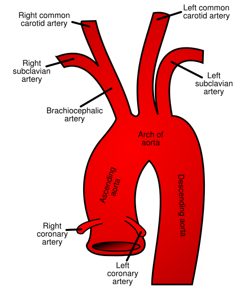
Image:Gray505.png|The arch of the aorta, and its branches. | Image:Gray505.png|The arch of the aorta, and its branches. | ||
Revision as of 17:28, 27 August 2013
|
WikiDoc Resources for Coronary arteries |
|
Articles |
|---|
|
Most recent articles on Coronary arteries Most cited articles on Coronary arteries |
|
Media |
|
Powerpoint slides on Coronary arteries |
|
Evidence Based Medicine |
|
Cochrane Collaboration on Coronary arteries |
|
Clinical Trials |
|
Ongoing Trials on Coronary arteries at Clinical Trials.gov Trial results on Coronary arteries Clinical Trials on Coronary arteries at Google
|
|
Guidelines / Policies / Govt |
|
US National Guidelines Clearinghouse on Coronary arteries NICE Guidance on Coronary arteries
|
|
Books |
|
News |
|
Commentary |
|
Definitions |
|
Patient Resources / Community |
|
Patient resources on Coronary arteries Discussion groups on Coronary arteries Patient Handouts on Coronary arteries Directions to Hospitals Treating Coronary arteries Risk calculators and risk factors for Coronary arteries
|
|
Healthcare Provider Resources |
|
Causes & Risk Factors for Coronary arteries |
|
Continuing Medical Education (CME) |
|
International |
|
|
|
Business |
|
Experimental / Informatics |
Editor-In-Chief: C. Michael Gibson, M.S., M.D. [1]; Rim Halaby, M.D. [2]; Hilda Mahmoudi M.D., M.P.H.[3]
Overview
The coronary arteries supply oxygenated blood to the heart muscle itself. Although blood fills the chambers of the heart, the muscle tissue of the heart, or myocardium, is so thick that it requires coronary blood vessels to deliver blood deep into it. There are two primary arteries supplying the heart, the left coronary artery and the right coronary artery. These two epicardial coronary arteries course along the surface of the heart and this is why they are called "epicardial" (on top of the heart) arteries. Smaller arteries dive deep into the heart muscle and are called subendocardial coronary arteries. The cardiac veins are the vessels that remove the deoxygenated blood from the heart muscle and return it to the right atrium.
Right Coronary Artery
- The right coronary artery (RCA) originates above the right cusp of the aortic valve. It travels down the right atrioventricular groove, towards the crux of the heart. In addition to supplying blood to the right ventricle (RV), the RCA supplies 25% to 35% of the left ventricle (LV).
- The right coronary artery branches into:
- The conus artery
- At the origin of the RCA is the conus artery.
- SA branch
- The RCA supplies the SA nodal artery in 60% of patients. The other 40% of the time, the SA nodal artery is supplied by the left circumflex artery.
- Acute marginal artery
- It supplies the right ventricular wall.
- Posterior descending artery
- In 85% of patients, the RCA gives off the posterior descending artery (PDA). In the other 15% of cases, the PDA is given off by the left circumflex artery.
- It supplies the inferior wall, posterior interventricular septum and posteromedial papillary muscle.[1]
- The conus artery
Additional Images
-

-
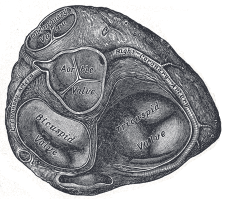
Base of ventricles exposed by removal of the atria.
-
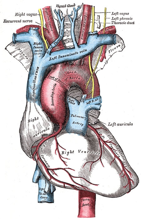
The arch of the aorta, and its branches.
-
Plan of the branches.
-
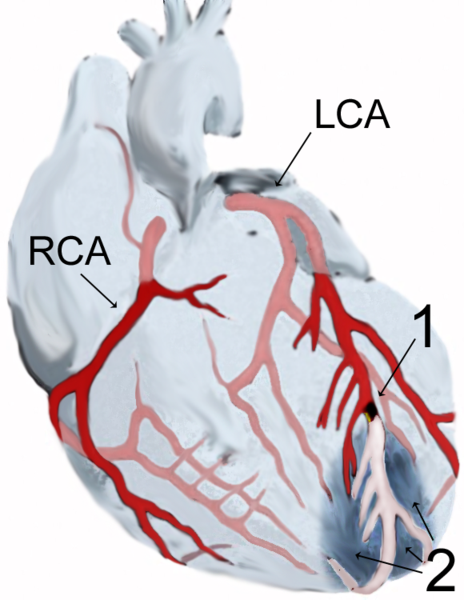
Diagram of a myocardial infarction.
-


Left Coronary Artery
The left coronary artery arises from the left aortic sinus. It bifurcates (divides or branches into) the Left anterior ascending artery (also known as the LAD or the anterior interventricular artery) and the Left circumflex artery (also known as the circumflex, circ, or LCX). It supplies the anterior part of the left ventricle: anterolateral myocardium, apex, anterior interventricular septum, anterolateral papillary muscle.
It supplies the posterolateral side of the left ventricle.
LCA with median ramus and marginal branches
File:LCA with median ramus.png
LCA with obtuse marginal branch

In order to objectively characterize the location of coronary obstructions, the following table is used to classify the coronary artery segments.
| Segment number | Segment label | Segment location | Segment discription | |
|---|---|---|---|---|
| 1 | R1 | Proximal right coronary artery | Extends from the ostium of the right coronary artery to the first of the three longest acute marginal branches. | |
| 2 | R2 | Mid right coronary artery | Extends from the origin of the first acute marginal branch to the origin of the third acute marginal branch. | |
| 3 | R3 | Distal right coronary artery | Extends from the origin of the third acute marginal to the origin of the posterior descending artery. | |
| 4 | R4 | Right posterolateral artery | This is the distal continuation of the right coronary artery after the origin of the posterior descending artery. It often has an inverted U shape as described by James and the AV nodal branch originates from this artery. It carries blood to the right posterior and right inferior arteries in large right dominant anatomy, to just the right inferior branch in small right dominant anatomy, and it is not present in left or balanced dominant systems. | |
| 5, 6, 7 | A1, A2, A3 | Acute marginal arteries | The longest three arteries arising from the right coronary artery to supply the right ventricular wall, numbered from proximal to distal. | |
| 8 | RD | Right posterior descending artery | In all but left dominant systems, this vessel runs in the posterior interventricular groove and supplies septal perforator branches. When present, it is one of the three longest branches on the inferior wall of the heart. | |
| 9 | RI | Right inferior artery | Arises from the fourth segment of the right coronary artery and supplies the inferior wall. In small right dominant anatomy, it is the distal most branch arising from the right coronary artery, while in large right dominant anatomy it arises proximal to the origin of the right posterior artery. When present, it is one of the three longest branches on the inferior wall of the heart. | |
| 10 | RP | Right posterior artery | Distal most branch to arise from the right coronary artery, but present only in large right dominant systems. When present, it is one of the three longest branches on the inferior wall of the heart. | |
| 11 | LM | Left main | Extends from the origin of the left coronary artery to the bifurcation into the left anterior descending and circumflex arteries. | |
| 12 | L1 | Proximal left anterior descending artery | Extends from the bifurcation of the left main coronary artery to the origin of the first septal artery. | |
| 13 | L2 | Mid left anterior descending artery | Extends from the origin of the first septal artery to the origin of the third septal artery. | |
| 14 | L3 | Distal left anterior descending artery | Extends from the origin of the third septal artery to the apex of the left ventricle. If there is no third septal branch, then the third segment begins halfway between S1 and the apex of the left ventricle. | |
| 15 | L4 | The left anterior descending artery terminus on the inferior wall | The continuation of the left anterior descending artery beyond the apex of the left ventricle in the event that the LAD is a wrap around variant. | |
| 16 | D1 | First diagonal artery | The first of the three longest branches off of the left anterior descending artery which supplies the anterolateral wall of the left ventricle. | |
| 17 | D2 | Second diagonal artery | The second of the three longest branches off of the left anterior descending artery which supplies the anterolateral wall of the left ventricle. In an RAO projection, this artery often arises where the left anterior descending angles toward the apex. | |
| 18 | D3 | Third diagonal artery | The third of the three longest branches off of the left anterior descending artery which supplies the anterolateral wall of the left ventricle. In an RAO projection, this artery often arises where the left anterior descending angles toward the apex. | |
| 19, 20, 21 | S1, S2, S3 | Septal arteries | The three largest branches off of the left anterior descending supplying the septum. | |
| 22 | MR | Median Ramus | An artery whose origin bisects the origins of both the left anterior descending artery and the circumflex artery. When a median ramus branch is present, the left main will be seen to trifurcate in the LAO caudal projection, and the median ramus artery is the middle artery at this point of trifurcation. This artery is also known as the intermedius. | |
| 23 | C1 | Proximal circumflex artery | Extends from the origin of the circumflex off of the left main to the origin of the first marginal or obtuse marginal branch. When a second obtuse marginal is present and the first marginal is absent, the C1- C2 transition is defined as halfway from the origin of the circumflex to the origin of the second obtuse marginal. | |
| 24 | C2 | Mid circumflex artery | Extends from the origin of the first marginal or obtuse marginal branch to the origin of the second marginal or obtuse marginal branch. When a second obtuse marginal is present and the first marginal is absent, the C1- C2 transition is defined as halfway from the origin of the circumflex to the origin of the second obtuse marginal. When a first obtuse marginal is present and the second marginal is absent, the C2- C3 transition is defined to be halfway from the first obtuse marginal to the end of C3. | |
| 25 | C3 | Distal circumflex artery | Extends from the origin of the second marginal or obtuse marginal to the termination of the circumflex artery in large right dominant anatomy or to the origin of the circumflex posterior branch (CP) in all other dominance. | |
| 26 | C4 | Left posterolateral artery | In left dominant or balanced systems this is the distal continuation of the circumflex artery in the atrio-ventricular groove. It carries blood to the left posterior descending artery and circumflex inferior artery in left dominant systems and to just the circumflex inferior artery in balanced dominant systems. | |
| 27 | CP | Circumflex posterior artery | In all but large right dominant anatomy, this branch originates at the distal end of the third segment of the circumflex at the border of the inferior and lateral left ventricular walls where it traditionally has been called a 4th marginal branch. When present, it is one of the three longest branches supplying the inferior wall of the heart. | |
| 28 | CI | Circumflex inferior artery | Arises from the fourth segment of the circumflex and supplies the inferior wall. In balanced dominant anatomy, it is the distal most branch arising from the circumflex, while in left dominant anatomy it arises proximal to the origin of the left posterior descending artery. When present, it is one of the three longest branches on the inferior wall of the heart. | |
| 29 | CD | Left posterior descending artery | In left dominant systems this is the distal continuation of the left circumflex artery which travels in the interventricular groove and supplies septal perforators at the base of the heart. This branch is the distal continuation of the circumflex after it leaves the atrio-ventricular groove in left dominant anatomy. When present, it is one of the three longest branches on the inferior wall of the heart. | |
| 30, 31, 32 | M1, M2, M3 | Marginal arteries | The three longest branches off of the circumflex artery supplying the lateral wall of the left ventricle, unless there is a large branching vessel which dominates the lateral left ventricular wall. When a large dominating artery is present, it is called an obtuse marginal. These marginal arteries are numbered from one (proximal) to three (distal). | |
| 33, 37, 41 | OM1-OM3 | Obtuse marginal arteries | A large branching artery which dominates the lateral left ventricular wall. An obtuse marginal is composed of anterior and posterior branches which share a common trunk. The anterior and posterior branches may substitute for the first and second marginal branches (or the second and third marginal branches), although a first (or third) marginal is permitted if present. No more than a single obtuse marginal may be present. An obtuse marginal is further specified as OM1 B OM3 depending on where the trunk arises compared to the usual origins of the first, second and third marginal arteries. | |
| 36, 40, 44 | OT | Obtuse marginal trunk | The common trunk of an artery connecting the anterior and posterior branches of an obtuse marginal to the circumflex. As no more than a single obtuse marginal may be present, the OT is not numbered. The 36 corresponds with OM1/OT, the 40 with OM2/OT, the 44 with OM3/OT | |
| 34, 38, 42 | OA | Anterior branch of the obtuse marginal artery | The anterior branch of an artery when it is a large branching vessel which dominates the lateral left ventricular wall. As no more than a single obtuse marginal may be present, the OA is not numbered. | |
| 35, 39, 43 | OP | Posterior branch of the obtuse marginal artery | The posterior branch of an artery when it is a large branching vessel which dominates the lateral left ventricular wall. As no more than a single obtuse marginal may be present, the OP is not numbered. The 35 corresponds with OM1/OP, the 39 with OM2/OP, the 43 with OM3/OP. | |
| 45 | SVGLAD | Saphenous Vein Graft to the LAD | ||
| 46 | SVGCX | Saphenous Vein Graft to Circumflex | ||
| 47 | SVGRCA | Saphenous Vein Graft to the Right Coronary Artery | ||
| 48 | SVGPDA | Saphenous Vein Graft to the PDA | ||
| 49 | SVGOM | Saphenous Vein Graft to the Obtuse Marginal | ||
| 50 | SVGD | Saphenous Vein Graft to Diagonal | ||
| 51 | LIMA | Left Internal Mammary Artery to the Left anterior descending artery |
Coronary Artery Dominance
- The dominance of coronary circulation is determined by the type of arteries that supply the posterior and inferior wall of the left ventricle.
- The artery that supplies the posterior descending artery (PDA) and the posterolateral artery (PLA) determines the coronary dominance.
- Right-dominant circulation: The right coronary artery (RCA) supplies both these arteries.
- Left-dominant circulation: The circumflex artery (CX), a branch of the left artery, supplies both these arteries.
- Co-dominant circulation: The RCA supplies the PDA and the CX supplies the PLA.
- Approximately 60% of the general population are right-dominant, 25% are co-dominant, and 15% are left-dominant.[2]
Blood Supply to the Papillary Muscles
- The papillary muscles tether the mitral valve (the valve between the left atrium and the left ventricle) and the tricuspid valve (the valve between the right atrium and the right ventricle) to the wall of the heart.
- If the papillary muscles are not functioning properly, the mitral valve leaks during contraction of the left ventricle and causes some of the blood to travel "in reverse", from the left ventricle to the left atrium, instead of forward to the aorta and the rest of the body.
- This leaking of blood to the left atrium is known as mitral regurgitation.
- The anterolateral papillary muscle:
- It receives two blood supplies: the LAD and LCX.
- It is therefore somewhat resistant to coronary ischemia (insufficiency of oxygen-rich blood).
- The posteromedial papillary muscle:
- It is supplied only by the PDA.
- This makes the posteromedial papillary muscle significantly more susceptible to ischemia.
- The clinical significance of the nature of blood supply to the papillary muscles is that a myocardial infarction involving the PDA is more likely to cause mitral regurgitation.
References
- ↑ Morton DA, Foreman KB, Albertine KH. Chapter 4. Heart. In: Morton DA, Foreman KB, Albertine KH, eds. The Big Picture: Gross Anatomy. New York: McGraw-Hill; 2011.
- ↑ Kaimkhani ZA, Ali MM, Faruqi AM (2005). "Pattern of coronary arterial distribution and its relation to coronary artery diameter". Journal of Ayub Medical College, Abbottabad : JAMC. 17 (1): 40–3. PMID 15929526.