Acute cholecystitis differential diagnosis: Difference between revisions
No edit summary |
No edit summary |
||
| Line 7: | Line 7: | ||
==Differentiating Acute cholecystitis from other Diseases== | ==Differentiating Acute cholecystitis from other Diseases== | ||
Acute cholecystitis must be differentiated from other diseases that cause right upper quadrant pain and nausea/vomiting such as:<ref name="BluthBenson2008">{{cite journal|last1=Bluth|first1=Edward I.|last2=Benson|first2=Carol B.|last3=Ralls|first3=Philip W.|last4=Siegel|first4=Marilyn J.|title=1: Right Upper Quadrant Pain|year=2008|doi=10.1055/b-0034-71418|url=https://www.thieme-connect.de/products/ebooks/lookinside/10.1055/b-0034-71418}}</ref><ref name="urlAcute cholecystitis | The BMJ">{{cite web |url=http://www.bmj.com/content/325/7365/639 |title=Acute cholecystitis | The BMJ |format= |work= |accessdate=}}</ref><ref name="pmid24679431">{{cite journal |vauthors=Knab LM, Boller AM, Mahvi DM |title=Cholecystitis |journal=Surg. Clin. North Am. |volume=94 |issue=2 |pages=455–70 |year=2014 |pmid=24679431 |doi=10.1016/j.suc.2014.01.005 |url=}}</ref> | Acute cholecystitis must be differentiated from other diseases that cause [[right upper quadrant pain]] and [[Nausea and vomiting|nausea/vomiting]] such as:<ref name="BluthBenson2008">{{cite journal|last1=Bluth|first1=Edward I.|last2=Benson|first2=Carol B.|last3=Ralls|first3=Philip W.|last4=Siegel|first4=Marilyn J.|title=1: Right Upper Quadrant Pain|year=2008|doi=10.1055/b-0034-71418|url=https://www.thieme-connect.de/products/ebooks/lookinside/10.1055/b-0034-71418}}</ref><ref name="urlAcute cholecystitis | The BMJ">{{cite web |url=http://www.bmj.com/content/325/7365/639 |title=Acute cholecystitis | The BMJ |format= |work= |accessdate=}}</ref><ref name="pmid24679431">{{cite journal |vauthors=Knab LM, Boller AM, Mahvi DM |title=Cholecystitis |journal=Surg. Clin. North Am. |volume=94 |issue=2 |pages=455–70 |year=2014 |pmid=24679431 |doi=10.1016/j.suc.2014.01.005 |url=}}</ref> | ||
*[[Biliary colic]] | *[[Biliary colic]] | ||
*[[Acute cholangitis]] | *[[Acute cholangitis]] | ||
| Line 546: | Line 546: | ||
{| | {| | ||
|- | |- | ||
| [[Image:Right_upper_quadrant.PNG|link=Right upper quadrant abdominal pain resident survival guide|339x339px]]|[[Image:Epigastric_quadrant_pain.PNG|link=Epigastric pain resident survival guide|179x179px]]|[[Image:Left_upper_quadrant.PNG|link=Left upper quadrant abdominal pain resident survival guide|329x329px]] | | <figure-inline>[[Image:Right_upper_quadrant.PNG|link=Right upper quadrant abdominal pain resident survival guide|339x339px]]</figure-inline><nowiki>|</nowiki><figure-inline>[[Image:Epigastric_quadrant_pain.PNG|link=Epigastric pain resident survival guide|179x179px]]</figure-inline><nowiki>|</nowiki><figure-inline>[[Image:Left_upper_quadrant.PNG|link=Left upper quadrant abdominal pain resident survival guide|329x329px]]</figure-inline> | ||
|- | |- | ||
|[[Image:Right_flank_quadrant.PNG|link=Right flank pain resident survival guide|338x338px]]|[[Image:Umbilical_pain.PNG|link=Umbilical region pain resident survival guide|165x165px]]|[[Image:Left_flank_quadrant.PNG|link=Left flank quadrant abdominal pain resident survival guide|335x335px]] | |<figure-inline>[[Image:Right_flank_quadrant.PNG|link=Right flank pain resident survival guide|338x338px]]</figure-inline><nowiki>|</nowiki><figure-inline>[[Image:Umbilical_pain.PNG|link=Umbilical region pain resident survival guide|165x165px]]</figure-inline><nowiki>|</nowiki><figure-inline>[[Image:Left_flank_quadrant.PNG|link=Left flank quadrant abdominal pain resident survival guide|335x335px]]</figure-inline> | ||
|- | |- | ||
|[[Image:Right_lower_quadrant.PNG|link=Right lower quadrant abdominal pain resident survival guide|338x338px]]|[[Image:Hypogastric.PNG|link=Hypogastric pain resident survival guide|199x199px]]|[[Image:Left_lower_quadrant.PNG|link=Left lower quadrant abdominal pain resident survival guide|335x335px]] | |<figure-inline>[[Image:Right_lower_quadrant.PNG|link=Right lower quadrant abdominal pain resident survival guide|338x338px]]</figure-inline><nowiki>|</nowiki><figure-inline>[[Image:Hypogastric.PNG|link=Hypogastric pain resident survival guide|199x199px]]</figure-inline><nowiki>|</nowiki><figure-inline>[[Image:Left_lower_quadrant.PNG|link=Left lower quadrant abdominal pain resident survival guide|335x335px]]</figure-inline> | ||
|} | |} | ||
Revision as of 23:21, 4 January 2018
|
Acute cholecystitis Microchapters |
|
Diagnosis |
|---|
|
Treatment |
|
Case Studies |
|
Acute cholecystitis differential diagnosis On the Web |
|
American Roentgen Ray Society Images of Acute cholecystitis differential diagnosis |
|
Risk calculators and risk factors for Acute cholecystitis differential diagnosis |
Editor-In-Chief: C. Michael Gibson, M.S., M.D. [1]; Associate Editor(s)-in-Chief: Furqan M M. M.B.B.S[2]
Overview
Acute cholecystitis must be differentiated from other diseases that cause right upper quadrant abdominal pain and nausea/vomiting such as biliary colic, acute cholangitis, viral hepatitis, alcoholic hepatitis, acute pancreatitis, acute appendicitis, and irritable bowel syndrome.
Differentiating Acute cholecystitis from other Diseases
Acute cholecystitis must be differentiated from other diseases that cause right upper quadrant pain and nausea/vomiting such as:[1][2][3]
- Biliary colic
- Acute cholangitis
- Viral hepatitis
- Alcoholic hepatitis
- Acute pancreatitis
- Acute appendicitis
- Irritable bowel syndrome
Abbreviations:
RUQ= Right upper quadrant of the abdomen, LUQ= Left upper quadrant, LLQ= Left lower quadrant, RLQ= Right lower quadrant, LFT= Liver function test, SIRS= Systemic inflammatory response syndrome, ERCP= Endoscopic retrograde cholangiopancreatography, IV= Intravenous, N= Normal, AMA= Anti mitochondrial antibodies, LDH= Lactate dehydrogenase, GI= Gastrointestinal, CXR= Chest X ray, IgA= Immunoglobulin A, IgG= Immunoglobulin G, IgM= Immunoglobulin M, CT= Computed tomography, PMN= Polymorphonuclear cells, ESR= Erythrocyte sedimentation rate, CRP= C-reactive protein, TS= Transferrin saturation, SF= Serum Ferritin, SMA= Superior mesenteric artery, SMV= Superior mesenteric vein, ECG= Electrocardiogram, US = Ultrasound
| |||||||||||||||||||||||||||||||||||||||||||||||||||||||||||||||||||||||||||||||||||||||||||||||||||||||||||||||||||||||||||||||||||||||||||||||||||||||||||||||||||||||||||||||||||||||||||||||||||||||||||||||||||||||||||||||||||||||||||||||||||||||||||||||||||||||||||||||||||||||||||||||||||||||||||||||||||||||||||||||||||||||||||||||||||||||||||||||||||||||||||||||||||||||||||||||||||||||||||||||
<figure-inline>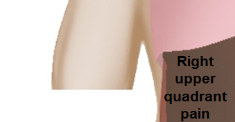 </figure-inline>|<figure-inline> </figure-inline>|<figure-inline> </figure-inline>|<figure-inline> </figure-inline>|<figure-inline> </figure-inline> </figure-inline>
|
<figure-inline> </figure-inline>|<figure-inline> </figure-inline>|<figure-inline> </figure-inline>|<figure-inline> </figure-inline>|<figure-inline> </figure-inline> </figure-inline>
|
<figure-inline> </figure-inline>|<figure-inline> </figure-inline>|<figure-inline> </figure-inline>|<figure-inline> </figure-inline>|<figure-inline> </figure-inline> </figure-inline>
|
References
- ↑ Bluth, Edward I.; Benson, Carol B.; Ralls, Philip W.; Siegel, Marilyn J. (2008). "1: Right Upper Quadrant Pain". doi:10.1055/b-0034-71418.
- ↑ "Acute cholecystitis | The BMJ".
- ↑ Knab LM, Boller AM, Mahvi DM (2014). "Cholecystitis". Surg. Clin. North Am. 94 (2): 455–70. doi:10.1016/j.suc.2014.01.005. PMID 24679431.