Sandbox:Sahar
| Lipomatous tumor | Age of onset | Gender preponderance | Location | Clinical features | Pathologic appearance | Other features | Pathologic view |
|---|---|---|---|---|---|---|---|
| Angiolipoma |
|
|
|
|
|
|
 |
| Myolipoma |
|
|
|
|
|
|
 |
| Myelolipoma |
|
|
|
|
|
|
 |
| Spindle Cell/Pleomorphic Lipoma |
|
|
|
|
|
|
 |
| Chondroid Lipoma |
|
|
|
|
|
|
 |
| Hibernoma |
|
|
|
|
|
|
 |
| Intramuscular and Intermuscular Lipomas |
|
|
|
|
|
|
 |
| Lipomas of Tendon Sheaths and Joints |
|
|
|
|
|
|
_ |
| Lumbosacral Lipoma |
|
|
|
|
|
|
_ |
| Neural Fibrolipoma (Lipofibromatous Hamartoma of Nerves) |
|
|
|
|
|
|
_ |
Example #1
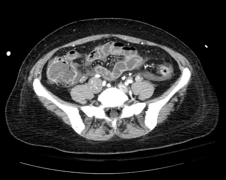
The patient presented with S.O.B. one year after hysterectomy for a leiomyomatous uterus.
-
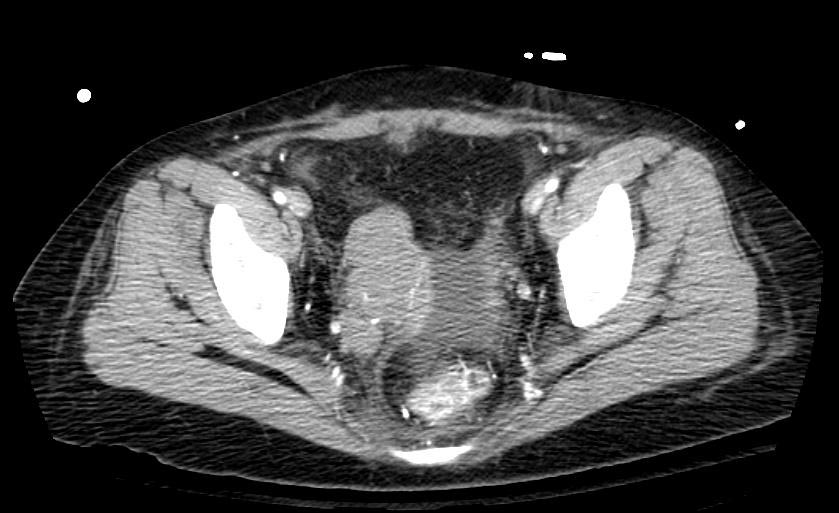
CT in Intravenous leiomyomatosis
-


- ↑ Stewart, Andrew F. (2005). "Hypercalcemia Associated with Cancer". New England Journal of Medicine. 352 (4): 373–379. doi:10.1056/NEJMcp042806. ISSN 0028-4793.
- ↑ Raftopoulos, Harry (2007). "Diagnosis and management of hyponatremia in cancer patients". Supportive Care in Cancer. 15 (12): 1341–1347. doi:10.1007/s00520-007-0309-9. ISSN 0941-4355.
- ↑ Barbosa, Sandrine Laboureau-Soares; Rodien, Patrice; Leboulleux, Sophie; Niccoli-Sire, Patricia; Kraimps, Jean-Louis; Caron, Philippe; Archambeaud-Mouveroux, Françoise; Conte-Devolx, Bernard; Rohmer, Vincent (2005). "Ectopic Adrenocorticotropic Hormone-Syndrome in Medullary Carcinoma of the Thyroid: A Retrospective Analysis and Review of the Literature". Thyroid. 15 (6): 618–623. doi:10.1089/thy.2005.15.618. ISSN 1050-7256.
- ↑ Florenzano, Pablo; Gafni, Rachel I.; Collins, Michael T. (2017). "Tumor-induced osteomalacia". Bone Reports. 7: 90–97. doi:10.1016/j.bonr.2017.09.002. ISSN 2352-1872.
- ↑ Nauck, Michael A.; Reinecke, Manfred; Perren, Aurel; Frystyk, Jan; Berishvili, Giorgi; Zwimpfer, Cornelia; Figge, Anja M.; Flyvbjerg, Allan; Lankisch, Paul G.; Blum, Werner F.; Klöppel, Günter; Schmiegel, Wolff; Zapf, Jürgen (2007). "Hypoglycemia due to Paraneoplastic Secretion of Insulin-Like Growth Factor-I in a Patient with Metastasizing Large-Cell Carcinoma of the Lung". The Journal of Clinical Endocrinology & Metabolism. 92 (5): 1600–1605. doi:10.1210/jc.2006-2573. ISSN 0021-972X.
- ↑ 6.0 6.1 Thiers, B. H.; Sahn, R. E.; Callen, J. P. (2009). "Cutaneous Manifestations of Internal Malignancy". CA: A Cancer Journal for Clinicians. 59 (2): 73–98. doi:10.3322/caac.20005. ISSN 0007-9235.
- ↑ Gaduputi, Vinaya; Chandrala, Chaitanya; Tariq, Hassan; Kanneganti, Kalyan (2014). "Sign of Leser-Trélat Associated with Esophageal Squamous Cell Cancer". Case Reports in Oncological Medicine. 2014: 1–3. doi:10.1155/2014/825929. ISSN 2090-6706.
- ↑ Tremblay, Catherine; Marcil, Isabelle (2017). "Necrolytic Migratory Erythema: A Forgotten Paraneoplastic Condition". Journal of Cutaneous Medicine and Surgery. 21 (6): 559–561. doi:10.1177/1203475417719051. ISSN 1203-4754.
- ↑ Cohen, Philip R.; Kurzrock, Razelle (1987). "Sweet's syndrome and malignancy". The American Journal of Medicine. 82 (6): 1220–1226. doi:10.1016/0002-9343(87)90229-4. ISSN 0002-9343.
- ↑ You, Hye Rin; Ju, Jae-Kyun; Yun, Sook Jung; Lee, Jee-Bum; Kim, Seong-Jin; Won, Young Ho; Lee, Seung-Chul (2018). "Paraneoplastic Pyoderma Gangrenosum Associated with Rectal Adenocarcinoma". Annals of Dermatology. 30 (1): 79. doi:10.5021/ad.2018.30.1.79. ISSN 1013-9087.
- ↑ 11.0 11.1 11.2 11.3 Pelosof, Lorraine C.; Gerber, David E. (2010). "Paraneoplastic Syndromes: An Approach to Diagnosis and Treatment". Mayo Clinic Proceedings. 85 (9): 838–854. doi:10.4065/mcp.2010.0099. ISSN 0025-6196.
- ↑ Abdulaziz, Ammar Taha Abdullah; Yu, Xiao Qing; Zhang, Le; Jiang, Xin Yue; Zhou, Dong; Li, Jin Mei (2018). "Paraneoplastic cerebellar degeneration associated with cerebellar hypermetabolism". Medicine. 97 (24): e10717. doi:10.1097/MD.0000000000010717. ISSN 0025-7974.
- ↑ Graus, F. (2001). "Anti-Hu-associated paraneoplastic encephalomyelitis: analysis of 200 patients". Brain. 124 (6): 1138–1148. doi:10.1093/brain/124.6.1138. ISSN 1460-2156.
- ↑ Shen, Kaini; Xu, Yan; Guan, Hongzhi; Zhong, Wei; Chen, Minjiang; Zhao, Jing; Li, Longyun; Wang, Mengzhao (2018). "Paraneoplastic limbic encephalitis associated with lung cancer". Scientific Reports. 8 (1). doi:10.1038/s41598-018-25294-y. ISSN 2045-2322.
- ↑ 15.0 15.1 Blaes, Franz (2013). "Paraneoplastic Brain Stem Encephalitis". Current Treatment Options in Neurology. 15 (2): 201–209. doi:10.1007/s11940-013-0221-1. ISSN 1092-8480.
- ↑ Gabrilovich, Mikhail; Raza, Mohammad; Dolan, Stephen; Raza, Tasleem (2006). "Paraneoplastic Polymyositis Associated With Squamous Cell Carcinoma of the Lung". Chest. 129 (6): 1721–1723. doi:10.1378/chest.129.6.1721. ISSN 0012-3692.
- ↑ "Paraneoplastic autonomic neuropathy: a case report | GM".
- ↑ Ascensao, Joad L.; Oken, Martin M.; Ewing, Stephen L.; Goldberg, Robert J.; Kaplan, Manuel E. (1987). "Leukocytosis and large cell lung cancer. A frequent association". Cancer. 60 (4): 903–905. doi:10.1002/1097-0142(19870815)60:4<903::AID-CNCR2820600431>3.0.CO;2-6. ISSN 0008-543X.
- ↑ Ahn, Heui June; Park, Yeon Hee; Chang, Yoon Hwan; Park, Sun Hoo; Kim, Min-Suk; Ryoo, Baek Yeol; Yang, Sung Hyun (2005). "A Case of Uterine Cervical Cancer Presenting with Granulocytosis". The Korean Journal of Internal Medicine. 20 (3): 247. doi:10.3904/kjim.2005.20.3.247. ISSN 1226-3303.
- ↑ Stone, Rebecca L.; Nick, Alpa M.; McNeish, Iain A.; Balkwill, Frances; Han, Hee Dong; Bottsford-Miller, Justin; Rupaimoole, Rajesha; Armaiz-Pena, Guillermo N.; Pecot, Chad V.; Coward, Jermaine; Deavers, Michael T.; Vasquez, Hernan G.; Urbauer, Diana; Landen, Charles N.; Hu, Wei; Gershenson, Hannah; Matsuo, Koji; Shahzad, Mian M.K.; King, Erin R.; Tekedereli, Ibrahim; Ozpolat, Bulent; Ahn, Edward H.; Bond, Virginia K.; Wang, Rui; Drew, Angela F.; Gushiken, Francisca; Lamkin, Donald; Collins, Katherine; DeGeest, Koen; Lutgendorf, Susan K.; Chiu, Wah; Lopez-Berestein, Gabriel; Afshar-Kharghan, Vahid; Sood, Anil K. (2012). "Paraneoplastic Thrombocytosis in Ovarian Cancer". New England Journal of Medicine. 366 (7): 610–618. doi:10.1056/NEJMoa1110352. ISSN 0028-4793.
- ↑ Rosu, Cristian; Cohen, Sandra; Meunier, Caroline; Ouellette, Denise; Beauchamp, Gilles; Rakovich, George (2011). "Pure red cell aplasia and associated thymoma". Clinics and Practice. 1 (1): 1. doi:10.4081/cp.2011.e1. ISSN 2039-7283.
- ↑ Leeaphorn, Napat; Kue-A-Pai, Pogsathorn; Thamcharoen, Natanong; Ungprasert, Patompong; Stokes, Michael B.; Knight, Eric L. (2014). "Prevalence of Cancer in Membranous Nephropathy: A Systematic Review and Meta-Analysis of Observational Studies". American Journal of Nephrology. 40 (1): 29–35. doi:10.1159/000364782. ISSN 1421-9670.