Whipworm infection pathophysiology
|
Whipworm infection Microchapters |
|
Diagnosis |
|---|
|
Treatment |
|
Case Studies |
|
Whipworm infection pathophysiology On the Web |
|
American Roentgen Ray Society Images of Whipworm infection pathophysiology |
|
Risk calculators and risk factors for Whipworm infection pathophysiology |
Editor-In-Chief: C. Michael Gibson, M.S., M.D. [1]
Overview
Infection is acquired by the ingestion of embryonated eggs from contaminated drinking water and food. The eggs once ingested hatch in the small intestine, and the larvae enter the intestinal crypts. The larve migrate to the proximal colon and mature into adult worms. The females begin to oviposit 60 to 70 days after infection and shed between 3,000 and 20,000 eggs per day. Whipworm causes disease by colonic mucosal invasion of the adult worms and resulting in inflammation of the colonic mucosa.
Pathophysiology
Life Cycle

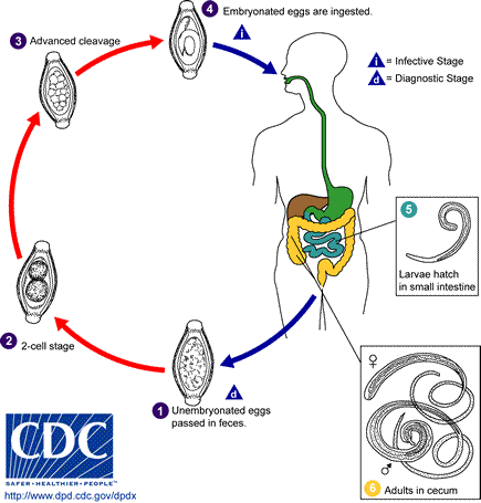
1.the eggs develop into a 2-cell stage 2.an advanced cleavage stage 3. and then they embryonate 4.eggs become infective in 15 to 30 days.5. mature and establish themselves as adults in the colon 6.The adult worms (approximately 4 cm in length) live in the cecum and ascending colon.The life span of the adult worm is about 1 year.
Transmission
Infection is acquired by the ingestion of embryonated eggs from contaminated drinking water and food.
Pathogenesis
- The eggs once ingested hatch in the small intestine, and the larvae enter the intestinal crypts.[1]*The larve migrate to the proximal colon and mature into adult worms.
- The adult worms live in the cecum and ascending colon and attach themselves to the colonic mucosa with the anterior portions threaded into the mucosa.
- The females begin to oviposit 60 to 70 days after infection and shed between 3,000 and 20,000 eggs per day.
- Whipworm causes disease by colonic mucosal invasion of the adult worms and resulting in inflammation of the colonic mucosa.
Associated Conditions
- Whipworm infection is frequently present in combination with Ascaris lumbricoides, hookworm and Entamoeba histolytica infections.
Gross Pathology
There are no specific gross pathology features associated with whipworm infection.
Microscopic Pathology
- Microscopy of the colonic mucosal biopsy from the affected site will demonstrate eosinophilia and neutrophilia.[2]
References
- ↑ Elston DM (2006). "What's eating you? Trichuris trichiura (human whipworm)". Cutis. 77 (2): 75–6. PMID 16570666.
- ↑ Kaur G, Raj SM, Naing NN (2002). "Trichuriasis: localized inflammatory responses in the colon". Southeast Asian J Trop Med Public Health. 33 (2): 224–8. PMID 12236416.