Superior vena cava syndrome chest x ray
Jump to navigation
Jump to search
|
Superior Vena Cava Syndrome Microchapters |
|
Differentiating Superior Vena Cava Syndrome from Other Diseases |
|---|
|
Diagnosis |
|
Treatment |
|
Case Studies |
|
Superior vena cava syndrome chest x ray On the Web |
|
American Roentgen Ray Society Images of Superior vena cava syndrome chest x ray |
|
Directions to Hospitals Treating Superior vena cava syndrome |
|
Risk calculators and risk factors for Superior vena cava syndrome chest x ray |
Editor-In-Chief: C. Michael Gibson, M.S., M.D. [1]Associate Editor(s)-in-Chief: Maria Fernanda Villarreal, M.D. [2]
Overview
On chest x-ray, indirect signs such as superior mediastinal widening and right hilar prominence may indicate the presence of a mediastinal mass.
Chest X Ray
On chest x-ray, indirect signs include:[1]
- Superior mediastinal widening
- Right hilar prominence that may indicate the presence of a mediastinal mass
Gallery
-
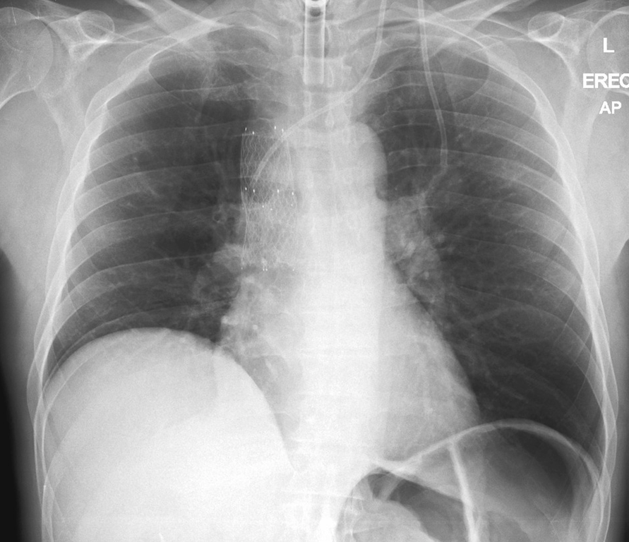 Superior mediastinal widening. Case courtesy of Dr Henry Knipe, Radiopaedia.org, rID: 31410
.png)
References
- ↑ Superior Vena Cava Syndrome.Dr Amir Rezaee and Radswiki et al. Radiopedia http://radiopaedia.org/articles/superior-vena-cava-obstruction Accessed on January 13, 2016