Parotid gland
Editor-In-Chief: C. Michael Gibson, M.S., M.D. [1]
Overview
|
WikiDoc Resources for Parotid gland |
|
Articles |
|---|
|
Most recent articles on Parotid gland Most cited articles on Parotid gland |
|
Media |
|
Powerpoint slides on Parotid gland |
|
Evidence Based Medicine |
|
Clinical Trials |
|
Ongoing Trials on Parotid gland at Clinical Trials.gov Trial results on Parotid gland Clinical Trials on Parotid gland at Google
|
|
Guidelines / Policies / Govt |
|
US National Guidelines Clearinghouse on Parotid gland NICE Guidance on Parotid gland
|
|
Books |
|
News |
|
Commentary |
|
Definitions |
|
Patient Resources / Community |
|
Patient resources on Parotid gland Discussion groups on Parotid gland Patient Handouts on Parotid gland Directions to Hospitals Treating Parotid gland Risk calculators and risk factors for Parotid gland
|
|
Healthcare Provider Resources |
|
Causes & Risk Factors for Parotid gland |
|
Continuing Medical Education (CME) |
|
International |
|
|
|
Business |
|
Experimental / Informatics |
The parotid gland is the largest of the salivary glands. It is found wrapped around the mandibular ramus, and it secretes saliva through Stensen's duct into the oral cavity, to facilitate mastication and swallowing.
Anatomy
Location
The parotid gland is found in the subcutaneous tissue of the face, overlying the mandibular ramus and anterior and inferior to the external ear. The gland occupies the parotid fascial space, an area posterior to the mandibular ramus, anterior and inferior to the ear. The gland extends irregularly from the zygomatic arch to the angle of the mandible. This gland is effectively palpated bilaterally. Start anterior to each ear and move to the cheek area and then inferior to the angle of the mandible. (Illustrated Head and Neck Anatomy, Fehrenbach and Herring, Elsevier, 2007, p. 170-1).
The facial nerve and its branches pass through the parotid gland as do the external carotid artery and its branches.
Excretory portion
The duct to this gland (also known as Stensen's duct) empties within the buccal cavity (the inside of the cheek) opposite the upper second molar. The parotid papilla is a small elevation of tissue that marks the opening of the parotid duct on the inner surface of the cheek (Illustrated Dental Embryology, Histology, and Anatomy, Bath-Balogh and Fehrenbach, Elsevier, 2006, p. 166).
Serous fluid (as opposed to mucous fluid) is produced by the parotid gland.
Innervation
Although the facial nerve (CN VII) runs through this gland, it does not control it.
Secretion of saliva by the parotid gland is controlled by presynaptic parasympathetic fibres originating in the inferior salivatory nucleus; these leave the brain via the glossopharyngeal nerve (CN IX), travel along the tympanic nerve (of Jacobson), pass through the tympanic plexus (located in the middle ear), and then travel in the lesser petrosal nerve until they reach the otic ganglion. After synapsing, the postganglionic fibers travel as part of the auriculotemporal nerve, a branch of the mandibular nerve (V3) to reach the parotid gland.
Sympathetic nerves originating from Superior Cervical Ganglion reach the gland with blood vessels.
Parasympathetic stimulation produces a water rich mucus saliva. Sympathetic stimulation leads to the production of low volume enzyme rich saliva. There is no inhibitory nerve supply to the gland.
Vascularization
Branches of the external carotid artery traverse the glandular tissue and supply the parotid gland with oxygenated blood, whereas numerous local veins drain the organ. These veins drain into tributaries of external and internal jugular veins.
The maxillary vein and superficial temporal vein meet to form the retromandibular vein within the parotid gland but are not responsible for draining it.
Lymphatics mainly comprise pre-auricular lymph nodes.
Pathology
Inflammation of one or both parotid glands is known as parotitis. The most common cause of parotitis was mumps. Widespread vaccination against mumps has markedly reduced the incidence of mumps parotitis. Other infections such as bacterial infections can cause parotitis as may blockage of the duct, whether from salivary duct calculi or external compression. Stones mainly occur within the main confluence of the ducts and within the main parotid duct. The patient usually complains of intense pain when salivating and tends to avoid foods which produce this symptom. In addition the parotid gland may become enlarged upon trying to eat. The pain can be reproduced in clinic via squirting lemon juice into the patient's mouth. Surgery depends upon the situation of the stone, if within the anterior aspect of the duct a simple incision into the buccal mucosa with sphinterotomy may allow removal, however if further posterior within the main duct, complete gland excision may be necessary.
The most common of tumors in the parotid gland are benign and only affect the superficial gland. These include pleomorphic adenoma and adenolymphoma. Their importance is in relation to their anatomical position. The tumorous growth can also change the consistency of the gland and cause facial pain on the involved side since the facial nerve travels through the gland (Illustrated Head and Neck Anatomy, Fehrenbach and Herring, Elsevier, 2007, p. 172). Critically, the relationship of the tumor to the branches of the facial nerve (CN VII) must be defined because resection may damage the nerves, resulting in paralysis of the muscles of facial expression. If the tumor is deep within the gland, the patient should give consent for potential damage of the facial nerve.
Additional images
-
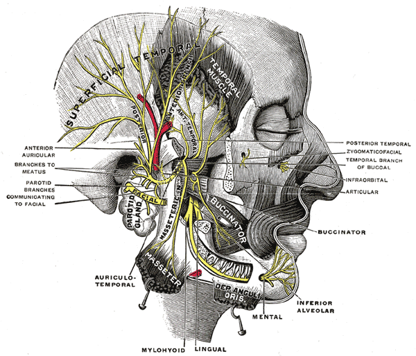 Mandibular division of the trifacial nerve.
 -
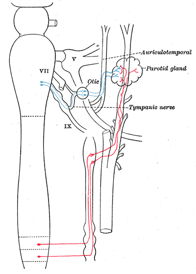 Sympathetic connections of the otic and superior cervical ganglia.
 -
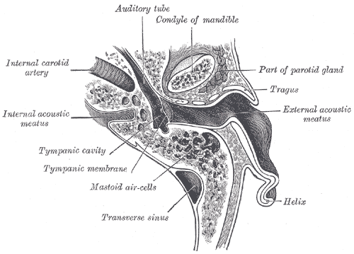 Horizontal section through left ear; upper half of section.
 -
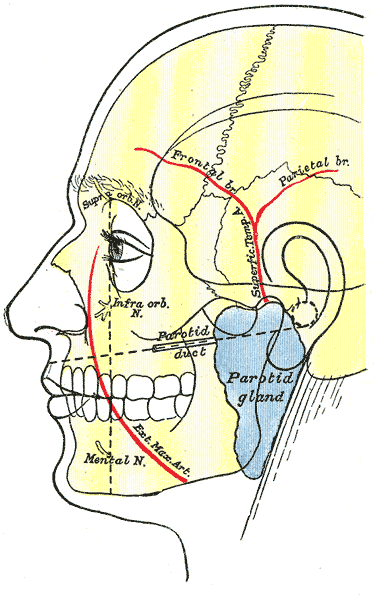 Outline of side of face, showing chief surface markings.
 -
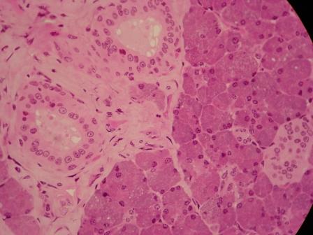 microscopic ducts surrounded by connective tissue


External links
- Illustration at yoursurgery.com
- Template:NormanAnatomy
- Histology at usc.edu
- Template:EMedicineDictionary
Template:Head and neck general
de:Ohrspeicheldrüse
it:Ghiandola parotide
sr:Доушна жлезда