Navicular bone
|
WikiDoc Resources for Navicular bone |
|
Articles |
|---|
|
Most recent articles on Navicular bone Most cited articles on Navicular bone |
|
Media |
|
Powerpoint slides on Navicular bone |
|
Evidence Based Medicine |
|
Clinical Trials |
|
Ongoing Trials on Navicular bone at Clinical Trials.gov Trial results on Navicular bone Clinical Trials on Navicular bone at Google
|
|
Guidelines / Policies / Govt |
|
US National Guidelines Clearinghouse on Navicular bone NICE Guidance on Navicular bone
|
|
Books |
|
News |
|
Commentary |
|
Definitions |
|
Patient Resources / Community |
|
Patient resources on Navicular bone Discussion groups on Navicular bone Patient Handouts on Navicular bone Directions to Hospitals Treating Navicular bone Risk calculators and risk factors for Navicular bone
|
|
Healthcare Provider Resources |
|
Causes & Risk Factors for Navicular bone |
|
Continuing Medical Education (CME) |
|
International |
|
|
|
Business |
|
Experimental / Informatics |
Editor-In-Chief: C. Michael Gibson, M.S., M.D. [1]
The navicular bone is one of the tarsal bones, found in the foot. Its name derives from the bone's resemblance to a small boat, caused by the strongly concave proximal articular surface. The term navicular bone or hand navicular bone was formerly used for the scaphoid bone, one of the carpal bones of the wrist.
It is located on the medial side of the foot, and articulates proximally with the talus, distally with the three cuneiform bones, and occasionally laterally with the cuboid.
As the bones in the foot develop, a particular process is important for the navicular structure. In the early stage, the navicular bone is actually cartilaginous and has to progress and calcify in order to maintain a strong form. There is a condition, however, in which the three cuneiforms that compose the bone do not completely calcify as a unit. In other words, they do not fuse properly, which leads to the most medial third creating a protrusion along the medial arch. This extension of the foot has a tendency to put stress on two tendons and the ligament that run along its side. The tendon of the peroneous brevis muscle which is the most distal of the two tendons, the tendon of the peroneous longus muscle which extends to the posterior of the ankle, and the posterior talofibular ligament which extends upward partway along the calf muscle, can all be potentially affected by this protrusion. Sometimes referred to as an extra navicular bone, this bump can cause wear and tear on the tendons causing sharp pain with increased activity. This condition can be corrected with surgery to file down or remove the protrusion and repair the tendons that were affected.
Resource
Names of the tendons and ligaments retrieved from Wexler, Randell K., M.D. “The Injured Ankle.” American Academy of Family Physicians. 1998 Vol. 57 No. 3. 7 February 2007 <http://www.aafp.org/afp/980201ap/wexler.html>.
See also
Additional images
-
 Bones of the right foot. Dorsal surface.
 -
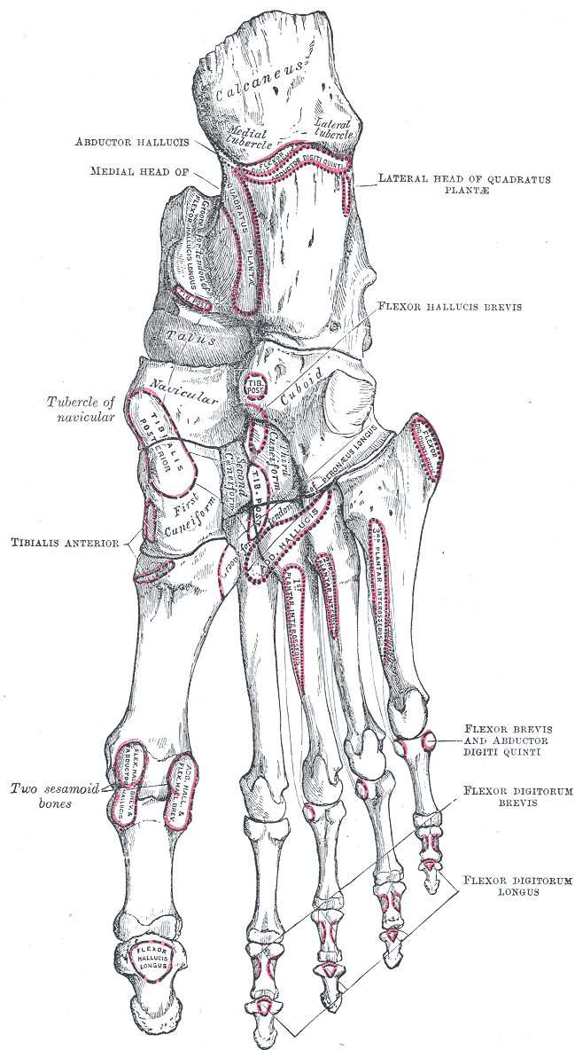 Bones of the right foot. Plantar surface.
 -
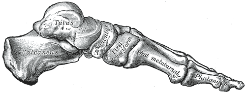 Skeleton of foot. Medial aspect.
 -
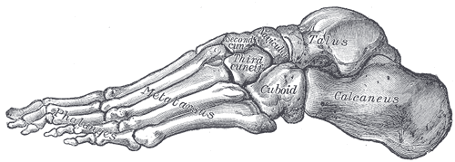 Skeleton of foot. Lateral aspect.
 -
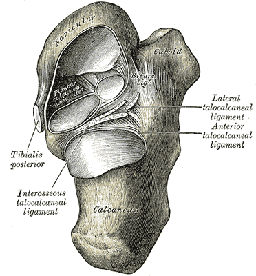 Talocalcaneal and talocalcaneonavicular articulations exposed from above by removing the talus.
 -
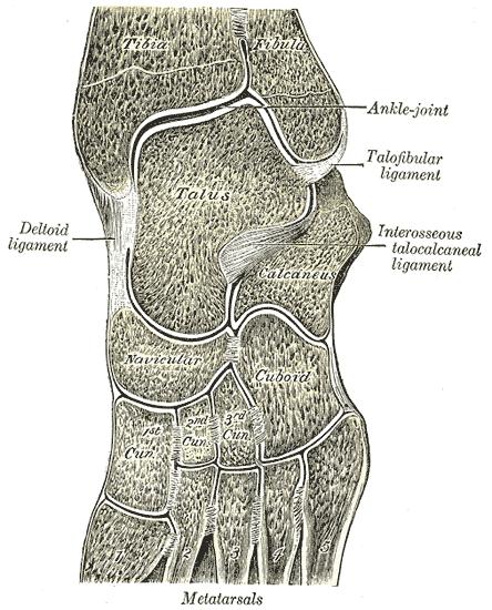 Oblique section of left intertarsal and tarsometatarsal articulations, showing the synovial cavities.


de:Os naviculare eu:Eskafoide (tarso) nl:Os naviculare no:Os naviculare sv:Båtben (fot) uk:Ладьєподібна кістка